Cavitation
What is Cavitation?
Cavitation refers to the nucleation, oscillation, and potential collapse of gas- or vapor-filled bubbles (cavities) in a liquid medium under the influence of oscillating acoustic pressure fields. In the context of ultrasound-tissue interaction, two regimes are classically distinguished:
- Inertial (transient) cavitation: bubbles expand rapidly and collapse violently, generating extreme local pressure, temperature, and shear forces. This is the dominant destructive mechanism in HIFU and is actively avoided in LIFU protocols (MI < 0.5).
- Stable (non-inertial) cavitation: bubbles oscillate periodically around an equilibrium radius without collapsing, producing sustained mechanical effects on surrounding structures. This is the regime assumed by all LIFU neuromodulation models.
The intramembrane cavitation hypothesis, first formalized by Krasovitski et al. (2011) in the NICE model, proposes a distinct third locus: cavitation occurring not in the bulk extracellular fluid but within the hydrophobic core of the lipid bilayer itself. The hypothesis posits that nanometer-scale gas pockets are pre-existing or nucleate between the two leaflets of the plasma membrane, forming a structure termed the bilayer sonophore (BLS). Under ultrasonic pressure oscillations, these intramembrane bubbles undergo stable oscillatory dynamics, causing periodic changes in membrane area, thickness, and capacitance that drive electrical currents through the membrane circuit.
Observed Effects and Predictions
The intramembrane cavitation framework predicts and, in computational models, reproduces a range of experimentally observed phenomena:
- Action potential generation at sub-millisecond timescales following ultrasound onset, consistent with electrophysiological recordings in cortical and peripheral neurons
- Threshold behavior in intensity and duty cycle: a minimum acoustic pressure is required to initiate sonophore oscillation, below which no membrane-level effect occurs
- Non-monotonic firing rate response: at very high intensities, excessive membrane oscillation drives depolarization block rather than sustained firing — reproduced in SONIC (Lemaire et al. 2019) and consistent with experimental observations by King et al. (2013, DOI: 10.1016/j.ultrasmedbio.2012.09.009)
- Cell-type selectivity: different neuron types respond differently to identical sonication parameters, arising from differences in resting membrane capacitance C_m0 — a direct prediction of the bilayer sonophore dynamics
- Frequency dependence: threshold intensity decreases as carrier frequency approaches the resonant frequency of the modeled sonophore, predicting optimal stimulation frequencies in the range of 0.3–1 MHz for CNS applications
Key Parameters and Modeling Conditions
The intramembrane cavitation framework introduces several biophysical parameters not present in standard HH models:
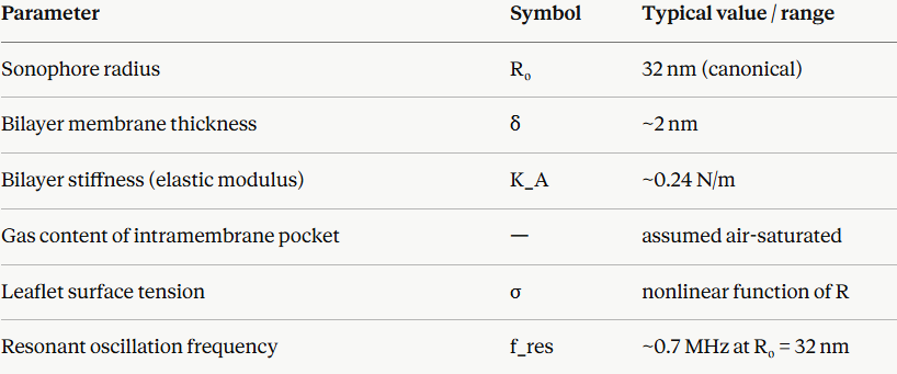
All NICE/SONIC-family models assume stable non-inertial oscillation of the bilayer sonophore, with the oscillation amplitude remaining well below the bubble collapse threshold. The models further assume that sonophore dynamics are spatially uniform across the membrane patch (point neuron assumption in NICE/SONIC, relaxed in MorphoSONIC).
Strengths of the Intramembrane Cavitation Approach
- Provides a complete mechanistic chain from acoustic pressure input to membrane electrical output, fully compatible with the Hodgkin-Huxley formalism
- Naturally accounts for cell-type selectivity through C_m0 differences without requiring additional free parameters
- Reproduces both excitatory and inhibitory (depolarization block - SONIC model) regimes within a single unified framework
- Computationally tractable via the lookup table coarse-graining approach introduced in SONIC, enabling simulation of realistic neuron morphologies and networks
- Predicts parameter-dependent thresholds consistent with experimental dose-response data
Limitations and Open Questions
The most fundamental limitation is that intramembrane cavitation has never been directly observed experimentally. The bilayer sonophore operates at nanometer spatial scales (32 nm radius) and nanosecond-to-microsecond temporal scales — both beyond the resolution limits of current experimental techniques, including fluorescence microscopy, AFM under sonication, and cryo-EM of sonicated membranes. The entire mechanistic chain from acoustic pressure to membrane oscillation therefore rests on theoretical and computational foundations without direct empirical confirmation.
Additional limitations include:
- Sonophore radius is a free parameter: R₀ = 32 nm is assumed rather than measured; model predictions are sensitive to this choice, and no method currently exists to determine the physiological distribution of sonophore sizes across neuron types
- Homogeneous membrane assumption: the bilayer is treated as a uniform elastic sheet, ignoring lipid raft heterogeneity, protein crowding, and cytoskeletal attachment — all of which would modulate local mechanical properties
- No interaction with MS channels in the base NICE/SONIC model: the cavitation-driven membrane tension that would activate TREK-1, TRAAK, or Piezo1 is not coupled back into the ionic current equations in the original framework (this coupling is addressed separately in the MS channel linking sub-model)
- Thermal effects are not included: NICE/SONIC models are purely mechanical; Q₁₀-dependent kinetic shifts from sonication-induced heating are absent from the base model
- Empirical status: should be treated as a theoretically motivated hypothesis, not established biophysical fact — a distinction important for interpreting model predictions