3' RNA Structures
SHAPE-map
(pmid: 32676598 Pyle 2020) The 3’ genomic terminus includes three well-studied stems, including the bulged-stem loop (BSL), Stem Loop 1 (SLI), and a long-bulge stem that includes the hypervariable-region (HVR), the S2M domain, the octanucleotide motif (ONM) subdomains, and a pseudoknot(Reviewed in (Yang and Leibowitz, 2015)). The consensus structure recapitulates the secondary structure of all the three stems with good SHAPE reactivity agreement
While the BSL is well determined in our structure, thelow reactivity for bulged nucleotides suggests the possibility of protein binding-partners (Fig2B, labeled BSL).
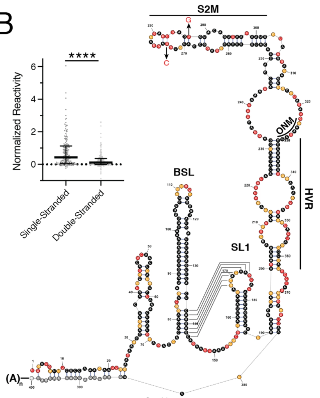
A pseudoknot structureis proposed toexist between the base of the BSL stem loop and the loop of SL1in coronaviruses(Yang and Leibowitz, 2015).While pseudoknot formation is mutually exclusive with the base of the BSL, studies in MHV have suggestedthat both structures contributeto viral replicationand themutually exclusive structuresare thought to function as a molecular switch in different steps of RNA synthesis(Goebel et al., 2004).
However, ourin-vivodetermined secondary structure is inconsistent withformation of the pseudoknot.
The low SHAPE reactivities for the nucleotides at the base of the BSL support formation of the extended BSL stem, while high-reactivities of the nucleotides in the loop of SLI indicate that it is highly accessible.Using theSHAPEKnots programfor robust predication of pseudoknots (implemented in RNA structure v5.8(Hajdin et al., 2013)), we found that apseudoknot is never predicted in three 500nt windows that cover the pseudoknotted region. Taken together, our data strongly support the extended BSL conformation, indicatingit is probably the dominant conformation in-vivo.
The HVR, so-named because it is poorly conserved across group II coronaviruses(Goebel et al., 2007),is predicted to be mostly single-stranded in our secondary structure, and the high reactivities across the span of this region lends strong experimental support for an unstructured region
We observe that thefirst three helices of S2M from SARS-CoV-2 exactly match the crystal structure determined for S2M from SARS-CoV (Robertson et al., 2005). However, our in-vivo structure deviates significantly at the top of the stem, with bases that are highly reactive
ONM is predicted at the central bulge between the S2M region and the HVR region. The sequence is absolutely conservedacross β-coronavirus(Goebel et al., 2007),but no functional significance has yet been shown.