Double membrane vesicles - DMV
DMVs
(Neufeldt 2020 doi: 10.1101/2020.06.23.167064) Similar to other (+)ssRNA viruses (7), coronaviruses modify cellular membranes to form double-membrane vesicles (DMVs), which are used as dedicated sites for vRNA replication (8). Several studies on βcoronaviruses SARS-CoV-1 and MERS-CoV that include electron tomography (ET) point towards DMVs being derived from ER cisternae in a process predominantly driven by the nonstructural protein 3 (nsp3) and nsp4 (9). In this model, the ER lumen constitutes the space between the DMV’s inner and outer membrane, while the enclosed space is of cytoplasmic origin and enriched in double-stranded RNA (dsRNA) (10).
Although the DMVs’ outer membranes are interconnected through narrow pores, the inner membrane appears to be sealed, posing the important question of how RNA is transported through the two membranes into the cytoplasm. Virion assembly takes place on the cytoplasmic side of the ER-Golgi intermediate compartment (ERGIC) cisternae (11).
DMVs might be a site of replication, it is as well possible that DMVs’ purpose is to conceal dsRNA transcription and replication byproducts that would otherwise be recognized by pattern recognition receptors of the innate immune system.
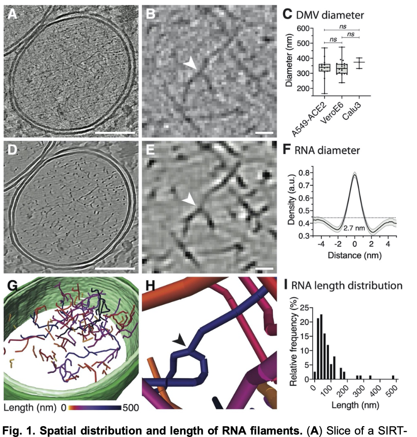
vRNA in the DMV
(Neufeldt 2020 doi: 10.1101/2020.06.23.167064) Individual vRNA filaments appeared smooth and did not organize into bundles or concentric layers
- uniform average diameter of 2.68 nm (SD = 0.23 nm, n = 80 from two tomograms) (Fig. 1F) and no electron-densities attributable to proteins attached to the fibers or to the inner DMV membrane were observed.
- observed filament branching points reminiscent of transcription intermediates representing a template that is copied with newly synthesized RNA being displaced from thetemplate
- Individual filaments featured a highly variable length ranging from 8 –487 nm with an average length of 76 nm (SD = 79 nm, n = 75). The length distribution corresponds to 29 –1739 base pairs taking into account the 0.28 nm rise per residue in the dsRNA A-helix (14). Considering that E and S, based on their gene length, would form 64 nm and 1070 nm long A-form helices, the observed filaments might represent dsRNA intermediates of subgenomic transcripts