Termination
Why does mRNA need a stop codon? When a ribosome reaches the codon UAG, UAA, or UGA on an mRNA strand then protein synthesis is terminated. A protein known as a release factor binds to the ribosome and adds a water molecule to the end of the amino acid chain. This reaction hydrolyzes the polypeptide chain from the tRNA, allowing the protein to exit the ribosome. Without the stop codon, protein synthesis would simply stop without releasing the completed protein.
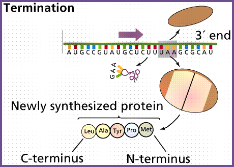
Termination occurs when the ribosome reaches a stop codon (UAA, UAG, and UGA). Since there are no tRNA molecules that can recognize these codons, the ribosome recognizes that translation is complete. The new protein is then released, and the translation complex comes apart.
Termination--Khan Academy Explanation--see opposite
Polypeptides, like all good things, must eventually come to an end. Translation ends in a process called termination. Termination happens when a stop codon in the mRNA (UAA, UAG, or UGA) enters the A site.
Stop codons are recognized by proteins called release factors, which fit neatly into the P site (though they aren't tRNAs). Release factors mess with the enzyme that normally forms peptide bonds: they make it add a water molecule to the last amino acid of the chain. This reaction separates the chain from the tRNA, and the newly made protein is released.
What next? Luckily, translation "equipment" is very reusable. After the small and large ribosomal subunits separate from the mRNA and from each other, each element can (and usually quickly does) take part in another round of translation.
Epilogue: Processing
Our polypeptide now has all its amino acids—does that mean it's ready to to its job in the cell?
Not necessarily. Polypeptides often need some "edits." During and after translation, amino acids may be chemically altered or removed. The new polypeptide will also fold into a distinct 3D structure, and may join with other polypeptides to make a multi-part protein.
Many proteins are good at folding on their own, but some need helpers ("chaperones") to keep them from sticking together incorrectly during the complex process of folding.
Some proteins also contain special amino acid sequences that direct them to certain parts of the cells. These sequences, often found close to the N- or C-terminus, can be thought of as the protein’s “train ticket” to its final destination. For more about how this works, see the article on protein targeting.

Colors: The large ribosome subunit is cyan, the small ribosome subunit pale yellow, EF-Tu red, and EF-G blue. tRNAs are gray (free or complexed with EF-Tu), magenta (binding at A site), green (in P site), yellow or brown (in the process of exiting).
Dr. Frank's laboratory group at the Wadsworth Center, New York State Department of Health, used cryo-EM and 3D image reconstruction to determine ribosomal structures and positions of EF-Tu and EF-G. Structures of EF-Tu and EF-G are based on separate X-ray crystallographic studies; https://www.rpi.edu/