Neutrophils
Return to Cardiovascular System Menu
Return to Immune System Menu
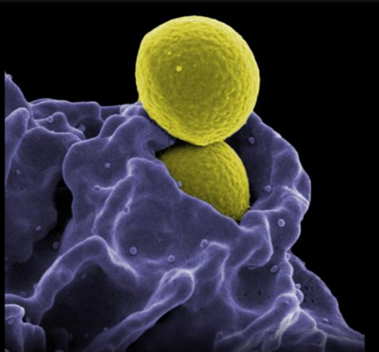
Image: Methicillin-resistant Staphylococcus aureus (MRSA) Ingestion by a Neutrophil.Scanning electron micrograph of MRSA bacteria (yellow) being ingested by a neutrophil (purple). (Credit: NIAID)
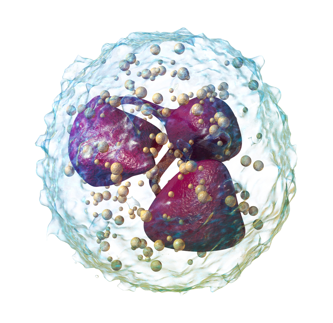
Comparison of Neutrophils to Monocytes
- Morphology: Neutrophils have a multi-lobed nucleus, while monocytes have a kidney-shaped or oval nucleus5.
- Lifespan: Neutrophils typically have a shorter lifespan (hours to days) compared to monocytes, which can survive longer and differentiate into macrophages or dendritic cells4.
- Timing of response: Neutrophils are usually the first to arrive at sites of inflammation, while monocytes arrive later6.
- Specialization: Neutrophils are more effective at rapid pathogen killing, while monocytes excel in antigen presentation and cytokine production8.
- Differentiation potential: Monocytes can differentiate into macrophages and dendritic cells, whereas neutrophils generally do not4.
While neutrophils and monocytes share some common features and work together in immune responses, they have distinct roles and characteristics that make them unique and complementary components of the immune system.
Neutrophils intentionally chew things up and spit them out in little pieces of antigens into the local fluids so other immune cells can pick them up and work with them. Or those broken pieces float all the way to the lymph node where there are immune cells to react to them.
Neutrophil Respiratory Burst
The neutrophil respiratory burst is a crucial immune response mechanism where neutrophils rapidly produce reactive oxygen species (ROS) to combat pathogens. This process is initiated when neutrophils encounter foreign invaders, leading to the activation of the enzyme NADPH oxidase. The enzyme catalyzes the reduction of oxygen to superoxide anion, which is further converted into other ROS like hydrogen peroxide and hydroxyl radicals. These reactive molecules are potent antimicrobial agents that help in destroying bacteria and fungi. However, excessive ROS production can also damage host tissues, highlighting the importance of regulating this response to prevent inflammatory diseases. Its believed Schisandra inhibits NADPH to reduce respiratory burst in neutrophils.
The polymorphonuclear leukocyte (PMN), or neutrophil, has long been recognized as the infantry of the innate immune system, rapidly deploying to sights of injury and infection. Considerable knowledge has accumulated demonstrating how these cells contribute to inflammation and host defense. In particular, the mechanisms of neutrophil recruitment, phagocytosis, nicotinamide adenine dinucleotide phosphate (NADPH) oxidative burst, and toxic granule-dependent microbial killing have been elucidated in great detail.
However, this conventional paradigm dramatically shifted with the observation that stimulated PMNs could release extracellular nucleic acids decorated with histones and granular proteins capable of entrapping exogenous bacteria. The discovery of these neutrophil extracellular traps (NETs) has spawned an entirely new field of granulocyte investigation. Since the initial description of NETs, as with most discoveries, more questions have been generated than answers. Source
Videos
Immune NETs: What COVID and Snake Venoms Have in Common
Neutrophil Mechanisms
Overview of Neutrophil Lineage
Hypersegmented
Neutrophils count; High or Low??? Know the Reasons
Neutrophil Contribution to Inflammation and Autoimmunity in Rheumatic Disease
How to raise neutrophils
These have a nucleus which is multi-lobed. Some have 2 or 3 or 4 lobes - which is why they're called "polymorphs".
The most abundant white cells are neutrophils and in a normal person the percentage is between 40-75%. If the neutrophil count is less than 40% its called neutropenia. If its over 75% its called neutrophilia (seen in acute inflammation). In acute inflammation many are released from bone marrow stores. These are heavily involved in any kind of infection that results in pus, such as strep or staph infections.
The most abundant type of granulocytes and most abundant (40%-70%) of white blood cells in most mammals. They form an essential part of the innate immune system.
They play an important role in acute inflammation, being the first to arrive in response to cell injury. They enter damaged tissue through a process called "neutrophil extravasation" in several distinct steps:
- Margination: when dilation of the blood vessels cause enough turbulence to force the neutrophils and other blood cells from the inner portion of the vessel to the peripheral portion where it starts bouncing into the wall.
- Rolling: the result of cytokines which cause the release of selectins on the surface of the endothelium cells lining the surface of the vessel. Selectins loosely interact with carbohydrates on the surface of the neutrophils causing it to sort of stick to the vessel wall so instead of bouncing along the vessel wall, it will roll due to loose interactions for a second , attach one part of the neutrophil to one part of the wall.
- Adhesion: additional cytokines cause the expression of CAMs (cellular adhesion molecules) on the surface of the endothelial cells of the vessel wall, as well as integrins on the surface of the neutrophil. This is a much tighter interaction than the rolling interaction, causing it to stop rolling and stick to the wall.
- Transmigration: This is when the neutrophil squeezes in between adjacent endothelial cells to get out of the vessel.
Once its through the vessel wall and into the tissue it will go on to migration towards the specific site of injury by being attracted to chemotactic factors (LTB4, C5a, IL-8)
They remove cell debris and are crucial for nerve damage. They used to think it was just macrophages that cleared debris but it turns out that neutrophils do the lion's share of that work.
How do monocytes and neutrophils interact during inflammation?
Monocytes and neutrophils engage in bidirectional cross-talk during inflammation, coordinating immune responses through physical interactions, cytokine signaling, and mutual recruitment. Here’s how their interplay unfolds:
Key Interaction Mechanisms
1. Reciprocal Recruitment
- Neutrophils recruit monocytes:
- Monocytes/macrophages recruit neutrophils:
2. Synergistic Activation
- Monocyte-driven neutrophil activation:
- Neutrophil-driven monocyte polarization:
3. Pathogen Clearance and Tissue Injury
- Cooperative phagocytosis:
- Amplified damage in dysregulation:
4. Regulatory Feedback
- Neutrophils limit monocyte hyperactivation:
- Monocytes regulate neutrophil lifespan:
Context-Dependent Roles
- In bacterial infections (e.g., Streptococcus suis):
- Neutrophils control bacterial burden, while monocytes exacerbate local inflammation in the CNS5.
- In sterile inflammation (e.g., glomerulonephritis):
- Monocyte–neutrophil interactions drive tissue injury via ROS and prolonged capillary retention1.
Clinical Implications
- Targeting monocyte–neutrophil interactions (e.g., blocking TNF-α or β2-integrins) may mitigate inflammatory damage in diseases like glomerulonephritis 1 6.
- Dysregulated cross-talk contributes to chronic inflammation in autoimmune and metabolic disorders 2 4.
This dynamic partnership balances pathogen clearance and tissue repair but can become destructive if unchecked, highlighting its dual role in health and disease.
What role does TNF play in the interactions between monocytes and neutrophils?
TNF-α serves as a central mediator of bidirectional communication between monocytes and neutrophils, orchestrating their recruitment, activation, and resolution during inflammation. Here’s how TNF-α shapes these interactions:
Recruitment and Adhesion
- Endothelial activation: TNF-α upregulates adhesion molecules (e.g., E-selectin, ICAM-1) on blood vessels, promoting neutrophil and monocyte adhesion and extravasation into tissues6.
- Chemokine induction: TNF-α stimulates monocytes to secrete CXCL1 and CXCL2, recruiting neutrophils, while neutrophils release azurocidin and LL-37 to attract monocytes16.
Activation and Cooperative Defense
- Monocyte-driven neutrophil priming:
- Neutrophil-driven monocyte polarization:
Resolution of Inflammation
- Efferocytosis regulation:
- Survival signals:
- Autonomous TNF-α signaling in monocytes activates NF-κB, preventing apoptosis and sustaining their presence during inflammation7.
Pathological Outcomes
- TNF-induced lethality: In sepsis, TNF-α triggers TRIF-dependent inflammatory cascades in neutrophils and apoptotic pathways in macrophages, driving organ failure4.
- Chronic inflammation: Dysregulated TNF-α loops (e.g., unresolved efferocytosis) perpetuate tissue damage in autoimmune diseases35.
Therapeutic Implications
- Etanercept (TNFR2-Fc fusion) blocks monocyte–tumor TNF-α/TNFR2 interactions, reversing protumorigenic signaling1.
- Targeting TRIF or CD14 pathways may mitigate TNF-driven sepsis lethality4.
By balancing proinflammatory and regulatory signals, TNF-α ensures coordinated monocyte–neutrophil responses but can become destructive if unchecked, highlighting its dual role in immune regulation.
PRESENCE IN GALT
Neutrophils are present in the gut-associated lymphoid tissue (GALT), particularly during inflammation. While GALT primarily consists of organized lymphoid structures such as Peyer's patches, isolated lymphoid follicles, and mesenteric lymph nodes, neutrophils are recruited to these areas under specific conditions. Here’s an overview of their role:
Presence and Recruitment of Neutrophils in GALT
- Recruitment During Inflammation:
- Neutrophils are recruited to the intestinal lamina propria and GALT in response to inflammatory signals such as chemokines (e.g., CXCL8/IL-8) and cytokines (e.g., TNF-α, IL-1β) produced by resident immune cells like macrophages and epithelial cells34.
- They traverse the vascular endothelium and can migrate into GALT-associated tissues to combat microbial invasion or injury3.
- Role in GALT:
- In Peyer's patches, neutrophils may contribute to the immune response by releasing antimicrobial peptides and cytokines that influence other immune cells, including B cells and T cells7.
- Neutrophils can also assist in antigen clearance and help regulate epithelial repair mechanisms during inflammation34.
Functional Roles in GALT
- Defense Against Pathogens: Neutrophils eliminate microbes that breach the epithelial barrier, protecting the gut from systemic infection 3.
- Regulation of Immune Responses: They produce cytokines like IL-22 and IL-17, which influence mucosal immunity and tissue repair 4.
- Interaction with Other Immune Cells: Neutrophils release chemoattractants (e.g., CAP18, cathepsin G) that recruit monocytes and macrophages for sustained inflammatory responses3.
Pathological Implications
Excessive neutrophil recruitment to GALT can result in tissue damage and contribute to conditions like inflammatory bowel disease (IBD), where their overactivation leads to mucosal injury and chronic inflammation 3 4.
In summary, while neutrophils are not a constitutive part of GALT under normal conditions, they play crucial roles in its function during inflammation by contributing to pathogen defense, immune regulation, and tissue repair.
Differences in neutrophil function within GALT compared to other lymphoid tissues
Neutrophils exhibit context-dependent functional adaptations in gut-associated lymphoid tissue (GALT) compared to other lymphoid tissues, shaped by the gut’s unique microbiota-rich environment. Here are the key differences:
Functional Specializations in GALT
1. Microbiota-Driven Modulation
- Balanced antimicrobial activity:
2. Antigen Handling and B Cell Interactions
- Support for IgA production:
- GALT neutrophils may indirectly aid IgA responses by clearing debris from germinal centers (GCs), allowing B cells to focus on glycan-specific IgA production against microbiota13.
- In non-GALT lymphoid tissues (e.g., lymph nodes), neutrophils instead enhance systemic antibody responses via direct antigen presentation to B cells26.
3. Tissue-Specific Recruitment and Positioning
- Proximity to epithelial barriers:
- GALT neutrophils patrol the follicle-associated epithelium (FAE), interacting with M cells and dendritic cells to sample luminal antigens14.
- In non-GALT lymphoid tissues, neutrophils localize to medullary cords or T cell zones, prioritizing interactions with adaptive immune cells over epithelial surveillance26.
4. Regulatory Mechanisms
- Enhanced tolerance induction:
5. Pathogen Clearance Strategies
- Cooperation with LysoDCs:
Comparative Roles in Lymphoid Tissues
| Feature | GALT Neutrophils | Non-GALT Lymphoid Tissue Neutrophils |
|---|---|---|
| Primary Trigger | Microbiota-derived signals | Pathogen invasion or sterile inflammation |
| Key Partners | LysoDCs, M cells, IgA⁺ B cells | T cells, follicular dendritic cells |
| Antibody Focus | IgA against bacterial glycans | IgG/IgM against protein antigens |
| Regulatory Role | Maintain microbiota tolerance | Amplify systemic adaptive responses |
| Tissue Damage Risk | Tightly controlled to protect epithelium | Higher potential for collateral damage |
Clinical Implications
- Dysregulated GALT neutrophil activity contributes to inflammatory bowel disease (IBD) due to breach of microbiota tolerance15.
- In systemic lymphoid tissues, neutrophil dysfunction is linked to autoimmune disorders (e.g., SLE) or impaired vaccine responses26.
Thus, GALT neutrophils are uniquely specialized to balance mucosal defense and microbiota coexistence, unlike their systemic counterparts, which prioritize pathogen clearance and adaptive immunity.
How does the gut microbiota influence neutrophil function in GALT?
The gut microbiota profoundly influences neutrophil function in gut-associated lymphoid tissue (GALT) through metabolic, inflammatory, and regulatory pathways. Here’s a synthesis of key mechanisms based on current evidence:
1. Regulation of Neutrophil Production and Recruitment
- Bone marrow myelopoiesis: Gut microbiota-derived signals (e.g., IL-17, G-CSF) modulate neutrophil production in the bone marrow. Depletion of microbiota reduces neutrophil numbers and impairs antimicrobial functions23.
- Chemokine signaling: Microbial metabolites like taurine (produced by Bacteroides uniformis) suppress NF-κB and IL-17 pathways, limiting excessive neutrophil recruitment to GALT during inflammation16.
2. Modulation of Neutrophil Activation and NETosis
- Suppression of NETs:
- Bacteroides uniformis and its metabolite taurine inhibit neutrophil extracellular traps (NETs) by deactivating IL-17 and NF-κB signaling, preventing pancreatic injury in hypertriglyceridemic pancreatitis (HTGP)16.
- Dysbiosis (e.g., reduced Bacteroides uniformis) disrupts this balance, leading to NET-driven tissue damage in GALT1.
- Pathogen defense: Commensal microbiota primes neutrophils for enhanced phagocytosis and ROS production via microbial components like peptidoglycan37.
3. Immune Crosstalk and Homeostasis
- IL-17/Th17 axis:
- Monocyte-neutrophil interactions: Microbiota metabolites (e.g., taurine) dampen monocyte-derived IL-17, reducing neutrophil hyperactivation and NETs in systemic circulation14.
4. Pathological Implications
- Inflammatory bowel disease (IBD): Dysbiosis disrupts neutrophil-microbiota crosstalk, leading to aberrant NETs, epithelial barrier damage, and chronic inflammation 6 7.
- HTGP: Loss of Bacteroides uniformis and taurine exacerbates NETs, driving pancreatic injury and systemic inflammation 1.
Therapeutic Opportunities
- Probiotics: Restoring Bacteroides uniformis or taurine supplementation reduces NETs and inflammation 1.
- Anti-IL-17 therapies: Targeting IL-17 signaling may mitigate neutrophil-mediated damage in GALT-associated disorders 1 6.
In summary, the gut microbiota fine-tunes neutrophil function in GALT through metabolic regulation, immune signaling, and suppression of pathological NETosis, maintaining a balance between mucosal defense and tolerance. Dysbiosis disrupts this equilibrium, driving inflammation and tissue injury.
IL-8 is a major chemotactic factor to recruit neutrophils. It also activates phagocytic cells.
Gliadin caused neutrophils to leave the capillaries and move into tissue. This happens independent of “leaky gut”. When the FPR1 receptor (which detects bacteria) was blocked, the neutrophils were also blocked.
Neutrophil activation can happen in some individuals when eating dairy casein. This is associated with food intolerance.
Streptococcal chemokine protease degrades the chemokine IL-8, which would otherwise attract neutrophils to the site of infection, so in a Streptococcus pyogenes infection be cautious about using agents that inhibit IL-8
Chronic Fatigue Syndrome: Mitochondrial dysfunction occurs in neutrophils in CFS patients . Neutrophils are the major effector cells of the immune system and the observed mitochondrial dysfunction is bound to have a deleterious effect on this system. Moreover, the degree of dysfunction was strongly correlated with the severity of their illness suggesting that the severity of a person’s CFS relates to the severity of the mitochondrial dysfunction.
Histamine: increases venule permeability, dilates the arterioles and from there, the vessel walls for neutrophil extravagation
ARTICLES/STUDIES
A better model for neutrophil-related diseases Neutrophils are critical immune cells for antimicrobial defense, but they can exacerbate a number of diseases, perhaps including COVID-19. The traditional approaches to study neutrophils in animal models are limited in specificity and effectiveness. Scientists have now identified the problem and have developed a new, optimized model for studying the role of neutrophils in the context of disease.
The intriguing role of polymorphonuclear neutrophils in antitumor reactions
Analysis Of Human Bone Marrow Reveals Neutrophil Progenitor
Neutrophil Extracellular Traps Awaken Sleeping Cancer Cells
Effects of ***Rosa canina** fruit extract on neutrophil respiratory burst
Arbutin** and decrease of potentially toxic substances generated in human blood neutrophils.
These results suggest that **Manuka honey** has an anti-inflammatory effect on neutrophils.
Rose hip** inhibits neutrophil respiratory burst.
Immunomodulatory activity of **triphala** on neutrophil functions.
Bromelain** treatment decreases neutrophil migration to sites of inflammation.
Bromelain** treatment decreases neutrophil migration to sites of inflammation.