H. Pylori Diagnostic Algorithm
Helicobacter pylori: An Update on Diagnostic Testing

Urea Breath Test - Helicobacter pylori - Infectious Peptic Ulcer.

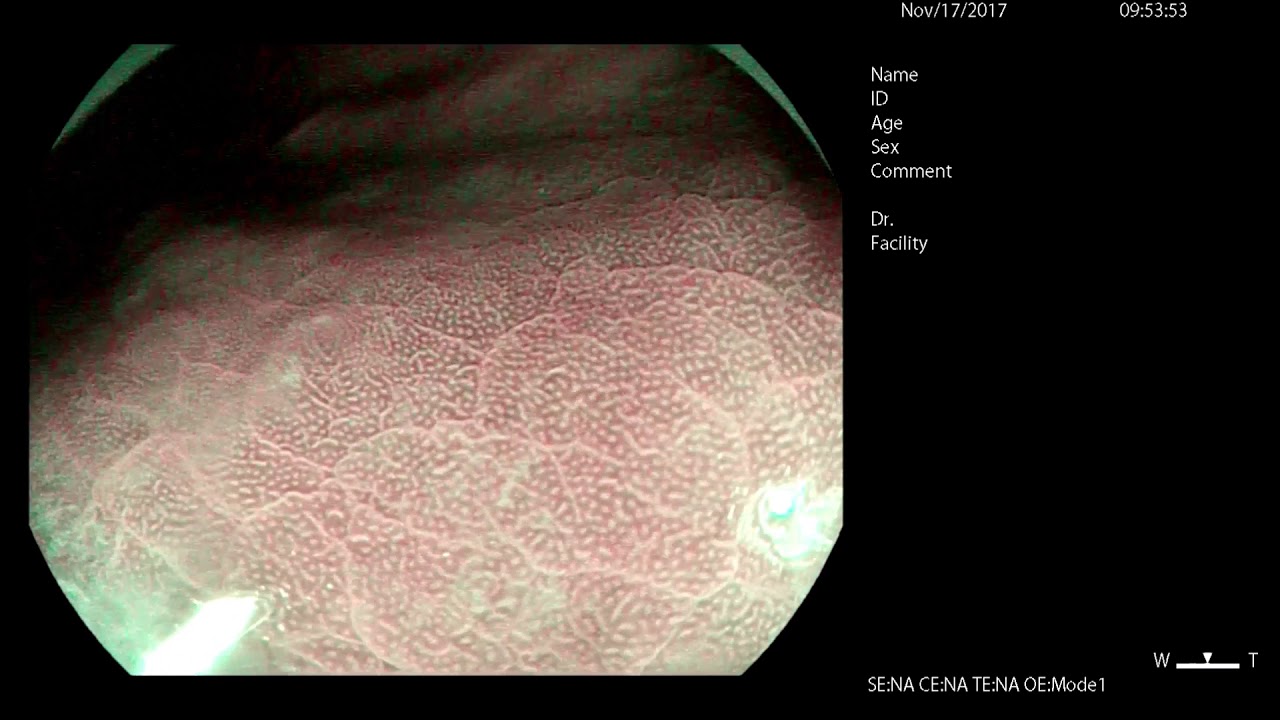
H. pylori Diagnosis with Magnifying Endoscopy
Helicobacter pylori (H. pylori) is a type of bacteria that infects the stomach lining and is a common cause of peptic ulcers. It is also associated with gastric cancer and mucosa-associated lymphoid tissue (MALT) lymphoma. Diagnosing H. pylori infection accurately is crucial for effective treatment and management. The H. pylori diagnostic algorithm is a systematic approach used by healthcare professionals to identify the presence of this bacterium in patients.
IMPORTANCE OF DIAGNOSING H. PYLORI
Diagnosing H. pylori is essential for several reasons:
- Treatment of Peptic Ulcers: Identifying H. pylori allows for targeted antibiotic therapy, which can lead to ulcer healing and prevent recurrence.
- Prevention of Gastric Cancer: Early detection and eradication of H. pylori can reduce the risk of developing gastric cancer.
- Symptom Relief: Proper diagnosis and treatment can alleviate symptoms such as abdominal pain, bloating, and nausea.
Diagnostic Methods
The H. pylori diagnostic algorithm includes both non-invasive and invasive methods. Each method has its own advantages and limitations, and the choice of test depends on the clinical scenario.
Non-Invasive Tests
- Urea Breath Test (UBT)
- Principle: Patients ingest a urea solution labeled with a carbon isotope. H. pylori produces urease, which breaks down urea, releasing labeled carbon dioxide that can be measured in the breath.
- Advantages: High sensitivity and specificity, non-invasive, and quick results.
- Limitations: Requires patient preparation, such as fasting and avoiding certain medications.
- Stool Antigen Test
- Principle: Detects H. pylori antigens in a stool sample.
- Advantages: Non-invasive, suitable for children, and can be used to confirm eradication after treatment.
- Limitations: Sensitivity may vary depending on the test kit used.
- Serology
- Principle: Detects antibodies against H. pylori in the blood.
- Advantages: Simple and widely available.
- Limitations: Cannot distinguish between current and past infections, as antibodies may persist after eradication.
Invasive Tests
- Endoscopy with Biopsy
- Principle: Direct visualization of the gastric mucosa and collection of biopsy samples for testing.
- Advantages: Allows for histological examination, culture, and rapid urease testing.
- Limitations: Invasive, requires sedation, and is more expensive.
- Rapid Urease Test (RUT)
- Principle: Biopsy samples are placed in a medium containing urea and a pH indicator. Urease activity from H. pylori changes the pH, causing a color change.
- Advantages: Quick results and high specificity.
- Limitations: False negatives can occur if the patient is on proton pump inhibitors (PPIs) or antibiotics.
The Diagnostic Algorithm
The H. pylori diagnostic algorithm is a step-by-step approach that guides clinicians in choosing the appropriate test based on the patient's clinical presentation and history.
Initial Assessment
- Evaluate Symptoms: Consider symptoms such as dyspepsia, peptic ulcer disease, or a family history of gastric cancer.
- Assess Risk Factors: Include factors like age, geographic location, and previous history of H. pylori infection.
Choosing the Right Test
- Non-Invasive Testing:
- Preferred for initial diagnosis in patients without alarm symptoms (e.g., weight loss, anemia, gastrointestinal bleeding).
- UBT or stool antigen test is recommended for most patients.
- Invasive Testing:
- Considered for patients with alarm symptoms or those undergoing endoscopy for other reasons.
- Endoscopy with biopsy and RUT are used to confirm the presence of H. pylori.
Post-Treatment Testing
- Confirm Eradication:
- Use UBT or stool antigen test at least four weeks after completing treatment to confirm eradication.
- Avoid serology for post-treatment testing due to persistent antibodies.