Acute Inflammation

Acute inflammation usually happens from minutes, to hours, to about 3 days.
In acute inflammation, 3 processes are going on at the same time:
- Active inflammation
- Tissue Injury largely produced by products of our own inflammatory cells, which can involve reactive oxygen species and other elements.
- Repair involves new blood vessel proliferation (angiogenesis) and fibrosis.
Mechanisms of acute inflammation consists of two phases involve with histamine, serotonin and kinin, released in the first hour (Ganesh et al., 2008) and with prostaglandin-like substances that are released in the second and third hours.
Causes of Acute Inflammation
- Infections (bacterial, viral, fungal, parasitic) – these are the most medically important
- Trauma (blunt and penetrating)
- Physical and chemical agents
- Tissue necrosis due to ischemia
- Immune reactions
- Foreign bodies (splinters, dirt, sutures)
It has 2 components:
Vascular Changes
- Vascular dilation: within tissues resident macrophages detect a pathogen, send chemical messages out to arteriole which then expands in diameter to increase blood flow. Inside the blood there are many inflammatory components. This increases hydrostatic pressure which drives fluid out between the cells of the arteriole into the tissue (transudate).
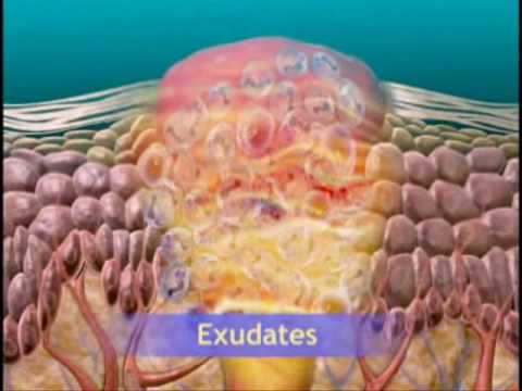
- Increase in vascular permeability: spaces open up between blood vessel cells to allow various immune defense components to leak out into the tissue cells. (exudate). The exudate consisting of a lot of fluid and protein is called edema, swelling at the site of the infection.
As proteins leak out, you increase concentration of red blood cells inside the vessel, increasing the viscosity (stasis) which slows down the flow of blood. This allows the white blood cells, primarily neutrophils, to move to the sides against the vessel wall (marginization). The fastest blood flow is in the center and it slows down towards the edge of the wall because there’s more friction. At this point, additional chemical changes allow the white blood cells to move through the gaps into the tissue.
There are several mechanisms that allow leukocytes to leave the blood stream into tissue:
- Endothelial Cell Concentration (most important first response) lasts 15-30 minutes. The endothelial cells lining the blood vessel wall contract.
- Cytoskeleton Changes: In the cell there are microfilaments that act as support structure in a 3-dimensional way. These shrink, and the cell gets smaller. TNF and IL-1 receptors are thought to cause these changes.
- Leukocyte Damage: As the leukocytes exit and respond to the emergency, they’re leaking all kinds of chemicals which can damage the endothelial cells surrounding the exit gap. There’s also a shear force as more and more exit which can also damage endothelial cells (which further promotes inflammation process).
Once transudate and exudate build up in the extracellular matrix, how does it get back into the veins? The lymphatic system runs parallel to the capillaries and it picks up all this stuff. Most of it will return into the vein side of the capillary but some will return into the lymph. The lymphatic system drains into nodes which are saturated with lymphocytes and other things. As the edema is drained from tissues that have an infection, the infection will drain down the lymphatic system. When red lines are seen streaking away from an infected cut, that’s a sign that the lymphatics are infected and if you then feel a big nodule on your neck, arm or chest its an inflammation of a lymph node, the infection has spread.
Cellular Events
The primary cellular event is increased number of neutrophils (polymorphonuclear leukocytes), these arrive first on the scene, the “first responder”.
Leukocyte Recruitment and Activation
Leukocytes are white blood cells responsible for killing the microbes, consuming bacteria, phagocytose (chew up) bad parts of cell debris and pathogens. They are responsible for cleaning up damaged tissue. So, its important that leukocytes are recruited and activated. But once they are activated, they can do damage to normal host cells which is why there’s a tightly regulated process by which they’re activated and recruited.
The steps:
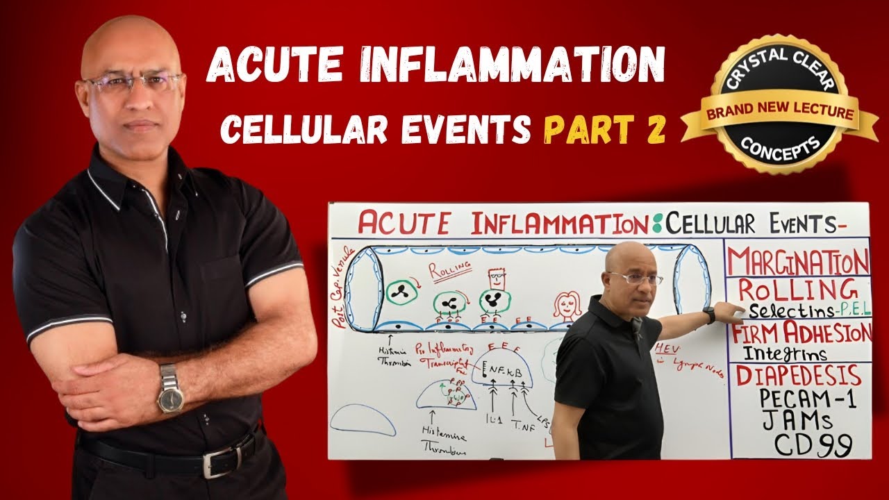
- Margination, adhesion to endothelium and rolling
- Firm adhesion to endothelium
- Transmigration between endothelium cells
- Migration in interstitial tissues towards a chemotactic stimulus
As blood slows down, white blood cells are larger than red blood cells so they’re naturally pushed to the side. Steps in recruitment start with margination. Rolling is the process by which it starts weakly attaching itself to selectin proteins, slowing it down until eventually it attaches firmly to these proteins. Once its stably attached it squeezed through endothelial cells. Once its outside the blood vessel its in the extracellular matrix.
Normally endothelial cells don’t have p-selectin and e-selectin on the surface, but when resident macrophages detect a problem and start destroying a pathogen they release cytokines (TNF, IL-1) that signal to express the selectins on outside of cell (they’re already IN the cell).
The chemokines are inflammatory markers secreted by macrophages that are already involved in the process. The chemokines go upwards toward the cell wall and signal the leukocytes in the blood stream to make a conformational change to their integrin receptor from a low affinity state to a high affinity state allowing it to change its shape allowing it to stop tolling and attach very strongly to an ICAM-1 molecule on the surface of the endothelial cell.
Once its completely stopped, the leukocyte expresses the PCAM-1 molecules and through diapedesis, comes out into the extracellular matrix. Then they undergo chemotaxis.
Chemotaxis
The white blood cell is like a police dog sniffing out its target. The bacteria is releasing it’s “stench” (bacterial products) into the extracellular matrix and its more concentrated right next to it. The process of degradation of any kind of cell or substance chemicals are released. On the white blood cell there are G-protein receptors which detect the concentration of these 4 types of chemicals, on the side that’s picking up a greater concentration, the cell will extend towards them creating an “arm” that attaches to the extracellular matrix filaments of fibrin and fibronectin, pulling itself towards the pathogen. Its using its calcium stores to help it do this. The “arms” are called pseudopods.
In the first 6-24 hours most of the cells are neutrophils and they only last this long. Days later you get monocytes, another type of white blood cell. There are more neutrophils in the blood and they make more adhesion molecules.
Inhibitory effect of anethole in nonimmune acute inflammation.