Atelectasis (lung collapse)
See also: Lung Disorders
Atelectasis - Resorption, Compression & Contraction
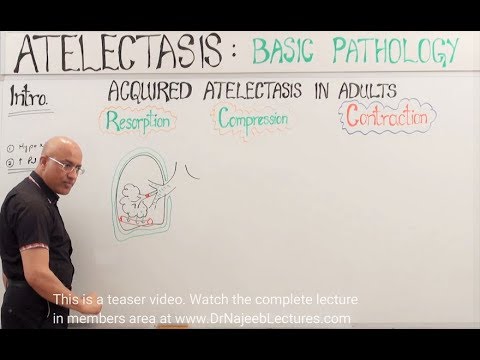
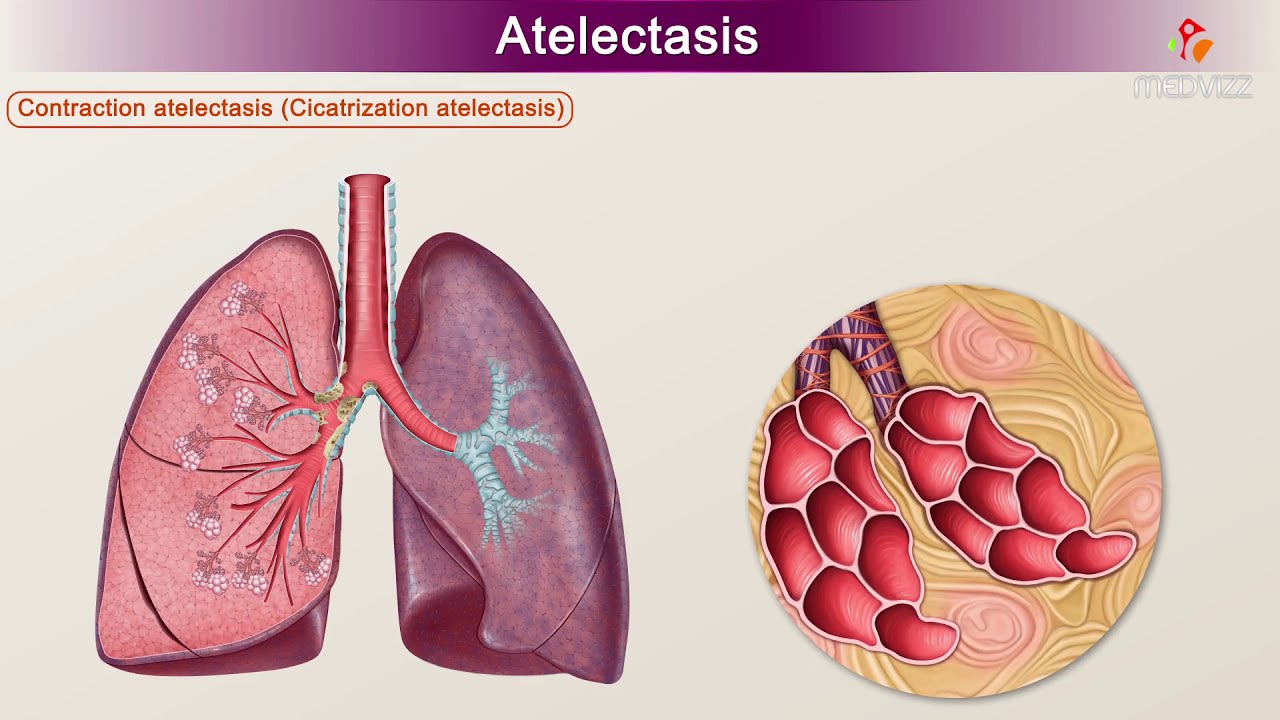
Atelectasis is a medical condition characterized by the partial or complete collapse of a lung or a section (lobe) of a lung. This condition occurs when the tiny air sacs (alveoli) within the lung become deflated or possibly filled with alveolar fluid. It is a common postoperative complication affecting the respiratory system, but it can also occur in a variety of other settings and due to multiple causes.
Causes of Atelectasis
Atelectasis can be caused by a variety of factors, including:
- Obstruction: This is one of the most common causes, where the airway is blocked by mucus, a foreign body, or a tumor, preventing air from reaching the alveoli.
- Compression: External pressure from the lung's surrounding structures, such as a pleural effusion (fluid in the space between the lungs and chest wall), pneumothorax, or tumor, can compress the lung.
- Postoperative Complications: Patients who have undergone surgery, especially abdominal or thoracic surgery, are at increased risk due to anesthesia, pain, and restricted breathing.
- Lung Conditions: Chronic lung diseases such as cystic fibrosis, COPD, or asthma can lead to atelectasis.
- Inactivity: Extended bed rest or lack of movement can lead to shallow breathing, which may result in atelectasis.
Symptoms of Atelectasis
Symptoms of atelectasis vary depending on the extent of lung tissue involved and may include:
- Difficulty breathing or shortness of breath.
- Rapid, shallow breathing.
- Coughing.
- Low-grade fever.
- Decreased breath sounds over the affected area.
- Possible cyanosis (bluish tint to the skin) in severe cases due to low oxygen levels.
Diagnosis
The diagnosis of atelectasis typically involves:
- Medical History and Physical Exam: A doctor will review the patient’s history and perform a physical examination focusing on the respiratory system.
- Imaging Tests:
* Chest X-ray: This is the most common method to confirm atelectasis, revealing areas of lung collapse.
* CT Scan: Provides a more detailed view and can help identify the cause of the collapse.
- Bronchoscopy: This procedure may be performed to look inside the airways, especially if an obstruction is suspected.
- Pulse Oximetry and Blood Gas Analysis: These tests assess the level of oxygen in the blood, which may be reduced in atelectasis.
Treatment and Management
The treatment of atelectasis focuses on re-expanding the collapsed lung tissue and may include:
- Chest Physiotherapy: Techniques like deep breathing exercises, coughing, and postural drainage can help remove blockages and improve lung expansion.
- Incentive Spirometry: This device encourages deep breathing to help re-expand the lung.
- Bronchodilators and Mucolytics: These medications can help open airways and thin mucus, respectively.
- Bronchoscopy: In cases of obstruction, bronchoscopy can be used to remove blockages like mucus plugs or foreign bodies.
- Surgery: Rarely, surgical intervention may be necessary to remove obstructions or correct anatomical issues.
Prevention
Preventive measures for atelectasis focus on maintaining clear airways and adequate lung expansion, especially in at-risk populations:
- Postoperative Care: Encouraging movement, deep breathing exercises, and the use of incentive spirometry can help prevent atelectasis after surgery.
- Regular Activity: Encouraging physical activity and avoiding prolonged bed rest can reduce the risk of lung collapse.
- Respiratory Therapy: For patients with chronic lung conditions, regular therapy can help maintain lung function.