Furin - S protein processing
Furin may cleave Protein S of SARS-CoV-2
Why did I go for Furin:(summary a little bit redundant but cites import work)
(Wrapp McLellan 2020 Science pmid: 32075877) 2019-nCoV S shares 98% sequence identity with the S protein from the bat coronavirus RaTG13, with the most notable variation arising from an insertion in the S1/S2 protease cleavage site that results in an “RRAR” furin recognition site in 2019-nCoV (19) rather than the single arginine in SARS-CoV (fig. S5) (20–23). Notably, amino acid insertions that create a polybasic furin site in a related position in hemagglutinin proteins are often found in highly virulent avian and human influenza viruses (24). In the structure reported here, the S1/S2 junction is in a disordered, solvent-exposed loop. In addition to this insertion of residues in the S1/S2 junction, 29 variant residues exist between 2019-nCoV S and RaTG13 S, with 17 of these positions mapping to the RBD (figs. S5 and S6). We also analyzed the 61 available 2019-nCoV S sequences in the Global Initiative on Sharing All Influenza Data database (https://www.gisaid. org/) and found that there were only nine amino acid substitutions among all deposited sequences.
(Galen Wo 2020 preprint) SARS-CoV-2 has a new Furin site - Using Bat CoV_RaTG13 Spike protein as reference (sharing 97% aa identity) the 4-amino acid insert can be identified as PRRA (AA position 681684). The effect of the 4-AA insertion is the presence of a furin signature sequence motif (PRRARSV) at the boundary of S1 and S2 domains of spike protein. This sequence motif consists the required Arg residue for P1 and P4 position of Furin site. In addition, it contains Arg at P3 site as well as Ser at P1’ site of furin motif. This sequence motif matches Aerolysin furin site in FurinDB and was predicted to be moderately strong (score 0.62) by ProP, a protease cleavage site prediction program. This finding suggests that the infectious 2019-nCoV virus, unlike SARS viruses, may be processed via cellular furin recognition and cleavage of the spike protein before host cell membrane fusion and entry.
(Coutard Decroly 2020 - pmid: 32057769) - Based on the 2019- nCoV S2′ sequence and the above arguments, we propose that one or more furin-like enzymes would cleave the S2′ site at KR↓SF.
Since furin is highly expressed in lungs, an enveloped virus that infects the respiratory tract may successfully exploit this convertase to activate its surface glycoprotein (Bassi et al., 2017; Mbikay et al., 1997). Before the emergence of the 2019-nCoV, this important feature was not observed in the lineage b of betacoronaviruses. However, it is shared by other CoV (HCoV-OC43, MERS-CoV, MHV-A59) harbouring furin-like cleavage sites in their S-protein (Fig. 2; Table 1), which were shown to be processed by furin experimentally (Le Coupanec et al.,2015; Mille and Whittaker, 2014). Strikingly, the 2019-nCoV S-protein sequence contains 12 additional nucleotides upstream of the single Arg↓ cleavage site 1 (Figs. 1B and 2) leading to a predictively solvent-exposed PRRAR↓SV sequence, which corresponds to a canonical furin- like cleavage site (Braun and Sauter, 2019; Izaguirre, 2019; Seidah and Prat, 2012). This furin-like cleavage site, is supposed to be cleaved during virus egress (Mille and Whittaker, 2014) for S-protein “priming” and may provide a gain-of-function to the 2019-nCoV for efficient spreading in the human population compared to other lineage b beta- coronaviruses. This possibly illustrates a convergent evolution pathway between unrelated CoVs. Interestingly, if this site is not processed, the S-protein is expected to be cleaved at site 2 during virus endocytosis, as observed for the SARS-CoV.
(Walls Veesler 2020 Cell - pmid: 32155444) SARS-CoV-2 S glycoprotein harbors a furin cleavage site at the boundary between the S1/S2 subunits, which is processed during biogenesis and sets this virusapart from SARS-CoV and SARS-related CoVs. cryo-EM structures of the SARS-CoV-2 S}:
Using western blot analysis, we observed that SARS-CoV-2 S was virtually entirely processed at the S1/S2 site during biosynthesis in HEK293T cells, presumably by furin in the Golgi compartment.We speculate that the almost ubiquitous expression of furin-like proteases could participate in expanding SARS-CoV-2 cell and tissue tropism, relative to SARS-CoV, as well as increasing its transmissibility and/or altering its pathogenicity.
Furin does not cleave Protein S of SARS-CoV
(Simmons Pöhlmann 2011 Virology - pmid: 21435673) - SARS-CoV
Take home message
It looks like the new Furin site is functional; corresponding to positions of highly virulent influenza viruses; the virus undergoes mutations in Protein S (therefore it is important to generate new tools)
Specific Furin Inhibitors may Stop virus spreading
see: Furin inhibitors - SARS-CoV-2
Reviews Corona-Furin
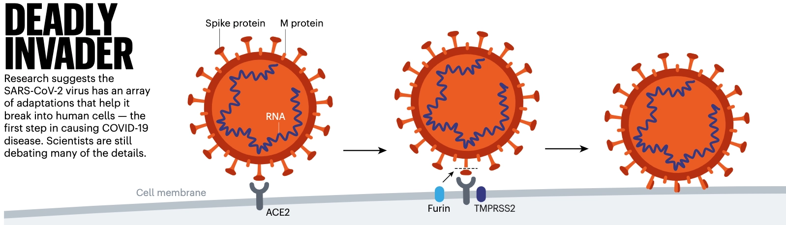
(Nature 7.May2020 p23 Profile of a Killervirus) SARS-CoV-2 is uniquely equipped for forcing entry into cells. Both SARS-CoV and SARS-CoV-2 bind with ACE2, but the receptor-binding domain of SARS-CoV-2 is a particularly snug fit. It is 10–20 times more likely to bind ACE2 than is SARS-CoV9. Wendtner says that SARS-CoV-2 is so good at infecting the upper respiratory tract that there might even be a second receptor that the virus could use to launch its attack. Even more troubling is the fact that SARS-COV-2 seems to make use of the enzyme furin from the host to cleave the viral spike protein. This is worrying, researchers say, because furin is abundant in the respiratory tract and found throughout the body. It is used by other formidable viruses, including HIV, influenza, dengue and Ebola to enter cells. By contrast, the cleavage molecules used by SARS-CoV are much less common and not as effective. Scientists think that the involvement of furin could explain why SARS-CoV-2 is so good at jumping from cell to cell, person to person and possibly animal to human. Robert Garry, a virologist at Tulane University in New Orleans, Louisiana, estimates that it gives SARS-CoV-2 a 100–1,000 times greater chance than SARS-CoV of getting deep into the lungs. “When I saw SARS-CoV-2 had that cleavage site, I did not sleep very well that night,” he says.