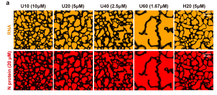
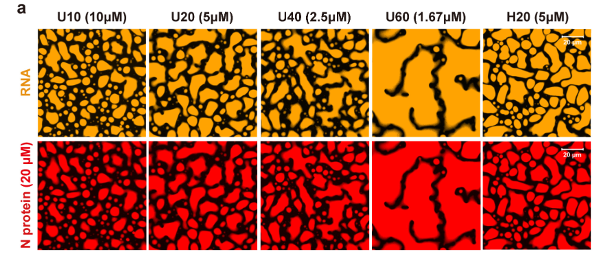
in vitro phase separation method - NP
(Chen Lou CellRes 2020 PMID: 32901111)
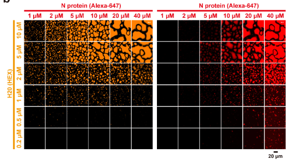
Imaging and data analysis of phase separation: Phase separation assays were performed in 1× PBS (pH 7.4) supplemented with 2.5% PEG8000 unless otherwise noted. A microwell 96-well optical-bottom plates with polymer base was coated with 1% BSA at 4 °C overnight and then washed with PBS three times. Phase separation was induced with RNA addition and then transferred in 96-well plate at room temperature for 1 h.
Imaging was performed with Olympus FV1200 equipped with a 60× oil immersion objective, and 3-6 regions were randomly selected for image acquisition. All images were processed with FV10-ASW, the percentage area of the image occupied by droplets was calculated based on the method from the published research 3.
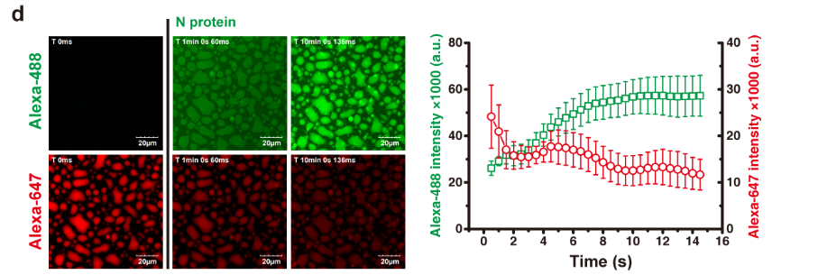
dynamics of droplets
length dependence of RNA