Spinal Reflexes
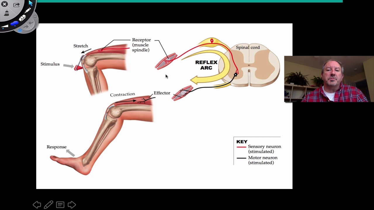
Spinal reflexes are fundamental components of the nervous system that allow for rapid and automatic responses to stimuli without the direct involvement of the brain. These reflexes are crucial for maintaining posture, protecting the body from harm, and facilitating everyday movements.
Anatomy of Spinal Reflexes
The spinal cord is a cylindrical structure that extends from the brainstem to the lower back, housed within the vertebral column. It serves as a conduit for transmitting neural signals between the brain and the rest of the body. The spinal cord is composed of gray matter, which contains neuron cell bodies, and white matter, which consists of myelinated axons. Reflex arcs, the neural pathways that mediate reflex actions, are primarily located within the gray matter of the spinal cord.
Components of a Reflex Arc
A typical reflex arc consists of five essential components:
- Receptor: A sensory organ that detects a specific stimulus.
- Sensory Neuron: Transmits the sensory information from the receptor to the spinal cord.
- Integration Center: Located within the spinal cord, where the sensory neuron synapses with a motor neuron or interneuron.
- Motor Neuron: Carries the response signal from the spinal cord to the effector.
- Effector: A muscle or gland that executes the response.
Types of Spinal Reflexes
Spinal reflexes can be categorized based on their complexity and the number of synapses involved:
Monosynaptic Reflexes
Monosynaptic reflexes involve a single synapse between the sensory and motor neurons. The most well-known example is the stretch reflex, such as the knee-jerk reflex (patellar reflex). When the patellar tendon is tapped, the quadriceps muscle is stretched, triggering a reflexive contraction to counteract the stretch.
Polysynaptic Reflexes
Polysynaptic reflexes involve one or more interneurons between the sensory and motor neurons, resulting in multiple synapses. These reflexes are more complex and can involve multiple muscle groups. An example is the withdrawal reflex, which occurs when a person touches a hot object. The reflex causes the immediate withdrawal of the hand to prevent injury.
Mechanisms of Spinal Reflexes
Spinal reflexes operate through a series of well-coordinated steps:
- Stimulus Detection: A receptor detects a stimulus, such as a change in muscle length or temperature.
- Signal Transmission: The sensory neuron transmits the signal to the spinal cord.
- Integration: The signal is processed in the spinal cord, where it may be relayed to interneurons for further processing.
- Response Generation: The motor neuron carries the response signal to the effector.
- Effector Action: The effector, typically a muscle, executes the response, such as contracting or relaxing.
Significance of Spinal Reflexes
Spinal reflexes play a vital role in maintaining homeostasis and ensuring survival. Their significance can be highlighted in several ways:
- Protection: Reflexes like the withdrawal reflex protect the body from harmful stimuli by enabling rapid responses without conscious thought.
- Posture and Balance: Reflexes help maintain posture and balance by adjusting muscle tension in response to changes in body position.
- Coordination: Reflexes contribute to the coordination of complex movements, such as walking, by ensuring that muscles work together efficiently.
- Diagnostic Tool: Reflex testing is a valuable diagnostic tool in clinical settings. Abnormal reflexes can indicate neurological disorders or damage to the nervous system.
Clinical Relevance
The assessment of spinal reflexes is a routine part of neurological examinations. Reflex tests can help identify issues such as:
- Hyperreflexia: Exaggerated reflexes, which may indicate upper motor neuron lesions or conditions like multiple sclerosis.
- Hyporeflexia: Diminished reflexes, which can suggest lower motor neuron damage or peripheral neuropathy.
- Areflexia: Absence of reflexes, often associated with severe nerve damage or spinal cord injury.