M cells / Microfold Cells
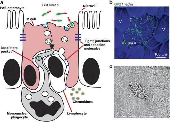
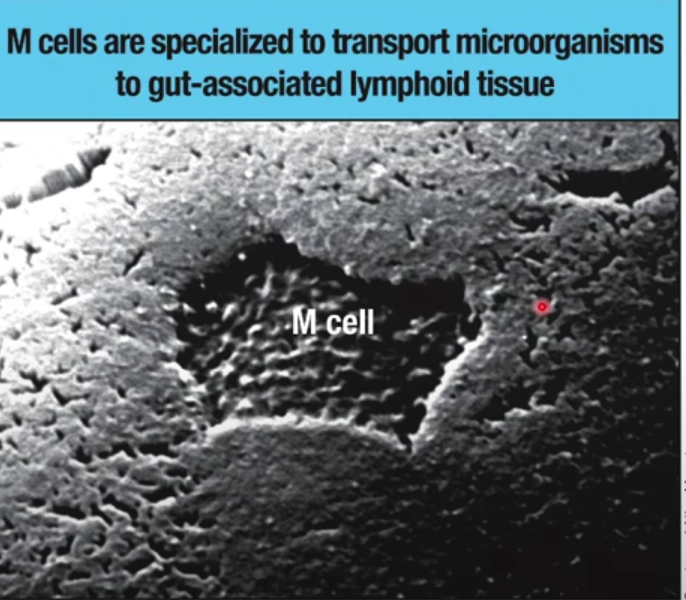
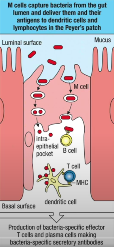
M-cells are specialized epithelial cells found in the gut-associated lymphoid tissue (GALT), primarily located in the follicle-associated epithelium of Peyer's patches in the small intestine 1 2 4. M cells are not only located in Peyer's patches; while they are most commonly found within the follicle-associated epithelium (FAE) of Peyer's patches, M cells can also be present in other areas of the gut-associated lymphoid tissue (GALT), like isolated lymphoid follicles and even in mucosal-associated lymphoid tissue (MALT) outside the gastrointestinal tract, such as in the nasopharynx. These unique cells play a crucial role in mucosal immune surveillance and have several distinctive characteristics:
Key Features
Morphology:
- Lack normal microvilli or have short, irregular microvilli
- Possess unique "microfolds" on their surface, which gives them their name
- Thin cytoplasm with a deeply invaginated basal membrane forming a pocket-like structure 3 4 6
Primary Functions
M-cells serve as gatekeepers of the mucosal immune system by:
- Sampling luminal microbes and antigens
- Transporting particles across the epithelial barrier through a process called transcytosis 1 2
- Delivering captured antigens to underlying immune cells like dendritic cells and lymphocytes 4 5
Transport Capabilities
They can transport various substances, including:
- Bacteria
- Viruses
- Parasites
- Non-infectious particles
- Macromolecules 5
Important Note: While M-cells are critical for immune surveillance, they can also be exploited by pathogens as an entry point for infection 5.
Location
M-cells are found in:
- Peyer's patches
- Intestinal lymphoid follicles
- Appendix
- Mucosal-associated lymphoid tissue sites
- Tonsils and adenoids 1 4 5
What Can Cause Damage to M-Cells?
Several factors can cause damage to M cells in the gut epithelium:
- Pathogenic bacteria: Certain enteropathogens can directly invade and destroy M cells, including:
- Salmonella typhimurium
- Salmonella typhi
- Shigella species
- Yersinia species
- Viral infections: Some viruses can exploit M cells as entry points, potentially causing damage:
- Prion infections: Prions can use M cells as a gateway, which may lead to cell damage 1.
- Inflammatory stimuli: Exposure to inflammatory factors can alter M cell density and potentially disrupt their normal function 1.
- Aging: The aging process can modify M cell density and potentially impact their integrity 1.
- Bacterial toxins: For example, botulinum toxin can enter through M cells, potentially causing harm 6.
- Rapid particle uptake: The specialized function of M cells in transcytosing particles can sometimes lead to their own destruction, especially when invaded by pathogens 3.
- Immune responses: In some cases, the immune response triggered by M cell-mediated antigen sampling can lead to local inflammation and potential damage to the cells themselves.
These factors can compromise the integrity of M cells, potentially disrupting their critical role in mucosal immunity and antigen sampling in the gut-associated lymphoid tissue (GALT).
Factors which impede M cells:
- Aging: The functional maturation of M cells is impeded by aging, which is associated with diminished antigen-specific immune responses 4.
- CCR6 deficiency: In CCR6-deficient mice, M-cell differentiation is impeded. The absence of CCR6 prevents the migration of certain cells towards the follicle-associated epithelium (FAE), which is necessary for M-cell maturation 4.
- RANKL blockade: Receptor activator of nuclear factor κB ligand (RANKL) is crucial for M-cell development. Selective deletion of membrane-bound RANKL in M-cell inducer cells abolishes the development of M cells in the follicle-associated epithelium 2.
- CSF1R signaling blockade: Depletion of CSF1R+CD68+ macrophages by blocking CSF1R signaling prominently decreases the expression of M-cell markers 2.
- Jagged1-Notch signaling: This pathway may regulate the M-cell population and/or maturation, potentially acting as a mechanism to prevent excessive M-cell formation 2.
These findings suggest that targeting these pathways or factors could potentially be used to modulate M-cell function, which could have implications for mucosal immunity and the development of mucosal vaccines.
What role does inflammation play in altering M cell density and function?
- Increased M cell density: During intestinal inflammation, there is a significant increase in the number of M cells, particularly in the colonic epithelium 1 2.
- Induction of Peyer's patch (PP) phenotype M cells: Inflammatory conditions lead to the development of PP-type M cells in the colon, often accompanied by loosely organized lamina propria infiltrates 2.
- Cytokine-driven M cell development: Inflammatory cytokines, particularly TNF-α and lymphotoxin-β receptor agonists, drive the differentiation of M cells 2.
- Increased apoptosis: Chronic intestinal inflammation is associated with an increase in M cell apoptosis, potentially compromising the epithelial barrie r3.
- Enhanced antigen uptake: M cell damage during inflammation appears to be responsible for increased uptake of microorganisms, potentially exacerbating the inflammatory response 3.
- Disruption of epithelial barrier: The increase in M cell number and function during inflammation can lead to a breakdown in the maintenance of the epithelial barrier 3.
- Potential role in pathogenesis: While M cells play a vital role in immune surveillance, their increased presence during inflammation may contribute to disease progression by allowing greater access to luminal antigens 2 3.
These alterations in M cell density and function during inflammation highlight the complex interplay between the immune system and the intestinal epithelium in maintaining gut homeostasis.
How do pathogens like Salmonella exploit M cells?
Pathogens like Salmonella have evolved sophisticated mechanisms to exploit M cells (microfold cells) for invasion and infection:
- Targeted entry: Salmonella typhimurium possesses a highly efficient mechanism for M cell entry, specifically targeting these cells in the follicle-associated epithelium (FAE )1.
- Membrane ruffling: S. typhimurium induces large membrane ruffles on the apical surface of M cells within 30-60 minutes of invasion, leading to cell necrosis and death 1.
- Type 3 Secretion System (T3SS): Salmonella uses a T3SS, a molecular syringe, to inject virulence effector proteins directly into M cells, manipulating host cell signaling pathways to force uptake 3.
- Exploitation of sampling function: Salmonella takes advantage of the M cells' natural role in sampling the intestinal lumen, using them as a route for invasion into the underlying mucosal lymphoid tissue 2 7.
- Transcription factor injection: Salmonella injects transcription factors into the gut to stimulate intestinal cells to become M cells, increasing potential entry points 6.
- Sugar binding: The bacterium attaches to sugars on the surface of M cells and uses its tentacles to prop open these cellular gates, facilitating entry into the intestine 6.
- Membrane reservoir exploitation: Salmonella exploits pre-existing tubular compartments associated with the plasma membrane, formed by RAB10 GTPase activity, to generate invasion ruffles 5.
- Immune system manipulation: After invasion, Salmonella can trigger the death of host macrophages and trick other immune cells into safely delivering them to new macrophages, evading destruction 4.
These diverse strategies demonstrate how Salmonella and other pathogens have adapted to efficiently exploit M cells as a primary route of invasion and infection in the intestinal epithelium.
Differences in how Salmonella and other pathogens like Shigella interact with M cells
- Invasion efficiency: Salmonella typhimurium possesses a highly efficient mechanism for M cell entry, specifically targeting these cells 1 2. In contrast, Listeria monocytogenes and Shigella flexneri appear to be internalized into M cells in a less disruptive fashion 1 2.
- Cell destruction: S. typhimurium's interaction with M cells is particularly dramatic. It induces large membrane ruffles on the apical surface of M cells within 30-60 minutes of invasion, leading to cell necrosis and death 1. Shigella and Listeria, while also causing cell death, do so in a less rapid and extensive manner 1 2.
- Invasion mechanism: Salmonella uses a Type 3 Secretion System (T3SS) to inject effector proteins into M cells, causing significant cytoskeletal rearrangements3. Shigella also uses a T3SS, but its interaction with M cells appears to be less disruptive initially 4.
- Post-invasion behavior: After invasion, Salmonella typically resides within a membrane-bound vacuole or freely within the host cell cytosol 5. Shigella, once in the cytosol, replicates and uses actin-based motility to escape host cell innate defenses and spread to neighboring cells 4.
- Immune response manipulation: Both Salmonella and Shigella manipulate the host immune response, but through different mechanisms. Salmonella can trigger the death of host macrophages, while Shigella uses its T3SS and effectors to block the host from mounting an efficient adaptive immune response 4.
These differences highlight the diverse strategies that enteric pathogens have evolved to exploit M cells as a route of invasion and infection in the intestinal epithelium.
How are M Cells replaced after Salmonella destruction?
M cells destroyed by bacteria are replaced through a dynamic process of intestinal epithelial regeneration involving several mechanisms:
- Lgr5+ intestinal stem cell (ISC) activation: Lgr5+ stem cells at the base of intestinal crypts rapidly respond to infection by increasing proliferation to replenish damaged epithelial cells, including M cells 1 2.
- Cellular remodeling: Infection triggers a rapid remodeling of the intestinal epithelium, with a decrease in stem cell proportions and an increase in progenitor cells, including transit amplifying (TA) cells, enterocyte progenitors, and Paneth cell progenitors 1.
- Dedifferentiation of progenitor cells: Various progenitor cells, such as label-retaining secretory precursors, Alpi+ enterocyte precursors, and Dll1+ progenitors, can dedifferentiate into actively cycling Lgr5+ stem cells to contribute to epithelial regeneration 2.
- Increased proliferation: Salmonella infection leads to enhanced stem cell and progenitor proliferation in intestinal crypts, as evidenced by increased crypt length and higher percentage of Ki67-positive cells 5.
- Activation of reserve stem cells: In cases of severe damage where Lgr5+ stem cells are lost, reserve stem cell populations can be activated to replenish the stem cell pool and aid in epithelial repair 6.
- Fetal-like conversion: Some differentiated cells, such as p57+ cells, undergo a dynamic reprogramming process after injury, characterized by a fetal-like conversion to support regeneration 4.
This multi-faceted regenerative response ensures the rapid replacement of damaged M cells and maintenance of the intestinal epithelial barrier function following Salmonella infection.
How quickly can intestinal stem cells regenerate M cells after Salmonella infection?
Intestinal stem cells can rapidly regenerate M cells and other epithelial cells after Salmonella infection through several mechanisms:
- Increased proliferation: Salmonella infection triggers enhanced stem cell and progenitor proliferation in intestinal crypts, evidenced by increased crypt length and a higher percentage of Ki67-positive cells 1 2.
- Rapid epithelial remodeling: Within 24 hours post-infection, there is a significant decrease in stem cell proportions and an increase in progenitor cells, including transit amplifying (TA) cells, enterocyte progenitors, and Paneth cell progenitors 1.
- Accelerated differentiation: Infected Lgr5+ intestinal stem cells (ISCs) rapidly differentiate into antimicrobial peptide (AMP)-secreting cells, mainly enterocytes and Paneth cells, as a defense mechanism against intracellular bacteria 1.
- Upregulated signaling pathways: Infection leads to elevated Wnt and Notch signaling in stem cells, which are important for Paneth and enterocyte lineage differentiation, respectively1.
- EGFR pathway activation: Both in vivo and organoid models show upregulation of EGFR-dependent signals, which are crucial for stem cell and progenitor proliferation and differentiation23.
While the exact timeframe for M cell regeneration is not explicitly stated, the intestinal epithelium's high turnover rate (normally 4-5 days) and the accelerated regeneration response to infection suggest that M cells can be replaced within a few days after Salmonella infection 5.
What role do Paneth cells play in the regeneration of M cells?
Paneth cells play a crucial role in the regeneration of intestinal epithelial cells, including M cells, through several mechanisms:
- Stem cell niche support: Paneth cells provide essential niche signals to neighboring intestinal stem cells (ISCs), which are responsible for replenishing all epithelial cell types, including M cells 1 4.
- Growth factor secretion: Paneth cells produce and secrete important growth factors such as EGF, Wnt3, and Notch ligands, which support Lgr5+ intestinal stem cells and promote their proliferation and differentiation 2 5.
- Wnt signaling: Paneth cells express canonical Wnt ligands (Wnt3a, Wnt9b, and Wnt11) that activate the Wnt signaling pathway in ISCs, driving β-catenin/Tcf signaling crucial for stem cell maintenance and differentiation1.
- Notch signaling: Paneth cells are a major source of Notch ligands DLL1 and DLL4, which bind to Notch receptors on ISCs and enterocyte progenitors, regulating their proliferation and differentiation 1.
- Regenerative capacity: Under stress conditions, Paneth cells can acquire stem cell features and directly contribute to intestinal regeneration, potentially including M cell replenishment 2 5.
- Microenvironment maintenance: By secreting antimicrobial peptides and regulating the intestinal microbiome, Paneth cells help maintain a healthy microenvironment conducive to proper epithelial regeneration 3.