Peptide-loading complex
MHC class I assembly and presentation

Assembly of the MHC I peptide-loading complex determined by a conserved ionic lock-switch
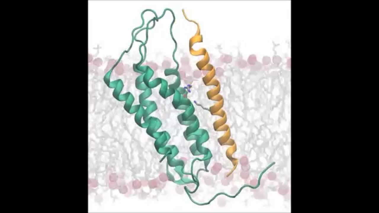
The peptide-loading complex (PLC) is a crucial component of the immune system, specifically involved in the presentation of antigens to T cells. This complex plays a vital role in the adaptive immune response by ensuring that peptides are properly loaded onto major histocompatibility complex class I (MHC I) molecules. Understanding the structure, function, and significance of the PLC is essential for comprehending how the immune system detects and responds to pathogens.
Structure of the Peptide-Loading Complex
The PLC is a multi-protein assembly that includes several key components:
- TAP (Transporter Associated with Antigen Processing): TAP1 and TAP2 form a heterodimer that transports peptides from the cytosol into the endoplasmic reticulum (ER).
- Tapasin: This chaperone protein bridges TAP and MHC I molecules, facilitating the transfer of peptides.
- ERp57: A thiol oxidoreductase that assists in the proper folding of MHC I molecules.
- Calreticulin: A chaperone that stabilizes MHC I molecules during peptide loading.
- MHC I molecules: These molecules present peptides on the cell surface for recognition by CD8+ T cells.
The assembly of these components ensures that peptides are efficiently loaded onto MHC I molecules, which is critical for the immune surveillance of infected or malignant cells.
Function of the Peptide-Loading Complex
The primary function of the PLC is to load peptides onto MHC I molecules. This process involves several steps:
- Peptide Generation: Proteins within the cell are degraded by the proteasome into peptide fragments.
- Peptide Transport: TAP transports these peptides from the cytosol into the ER.
- Peptide Editing: Tapasin, along with ERp57 and calreticulin, ensures that only high-affinity peptides are loaded onto MHC I molecules.
- MHC I Stabilization: The binding of a high-affinity peptide stabilizes the MHC I molecule, allowing it to be transported to the cell surface.
- Antigen Presentation: Once on the cell surface, the peptide-MHC I complex can be recognized by CD8+ T cells, initiating an immune response.
Significance in Immune Response
The PLC is essential for the immune system's ability to detect and respond to intracellular pathogens, such as viruses and some bacteria, as well as cancer cells. By presenting peptides derived from these pathogens or abnormal cells, the PLC enables the immune system to:
- Identify Infected Cells: CD8+ T cells can recognize and kill cells presenting foreign peptides.
- Eliminate Cancer Cells: Tumor antigens presented by MHC I molecules can be targeted by the immune system.
- Maintain Immune Surveillance: Continuous monitoring of cellular health and the presence of abnormal proteins.
Clinical Implications
Defects in the PLC can lead to severe immunodeficiencies and increased susceptibility to infections and cancer. Understanding the PLC has significant clinical implications:
- Immunotherapy: Enhancing the function of the PLC can improve the efficacy of cancer immunotherapies.
- Vaccine Development: Vaccines that target the PLC can enhance the presentation of antigens and improve immune responses.
- Autoimmune Diseases: Dysregulation of the PLC can contribute to autoimmune diseases, where the immune system mistakenly targets healthy cells.
Research and Future Directions
Ongoing research aims to further elucidate the mechanisms of the PLC and its components. Key areas of interest include:
- Structural Studies: High-resolution structures of the PLC components and their interactions.
- Regulation: Understanding how the PLC is regulated under different physiological conditions.
- Therapeutic Targets: Identifying new targets within the PLC for therapeutic intervention.
Advancements in these areas could lead to novel treatments for a variety of diseases, including infections, cancer, and autoimmune disorders.
The major histocompatibility complex class-I (MHC-I) peptide-loading complex (PLC) translocates cytosolic degradation products to the endoplasmic reticulum to load antigenic peptides onto MHC-I molecules. Stable peptide–MHC-I complexes are presented at the cell surface to mirror cellular contents for patrolling T cells, which protect against viral infections and cancer-causing mutations by inducing apoptosis in cells that expose nonself peptides. Due to its size and dynamic nature, the atomic-level details of the PLC remained unknown. We built an all-atom model of the human MHC-I PLC by combining the recent 9.9-Å resolution cryo-EM density with microsecond molecular dynamics simulations in a membrane and water environment (1.6 million atoms). The results provide unprecedented insights into the molecular underpinnings of our adaptive immune response. Study
Antigen Presentation: Visualizing the MHC Class I Peptide-Loading Bottleneck