Differentiating Shock
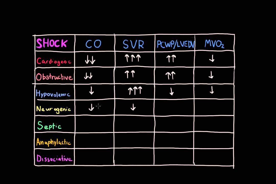
Understanding and Differentiating Shock
Shock is a critical medical condition characterized by the inadequate delivery of oxygen and nutrients to the tissues, leading to cellular and organ dysfunction. It is a life-threatening state that requires prompt recognition and treatment. Differentiating between the various types of shock is crucial for effective management and improving patient outcomes. This note explores the different types of shock, their causes, clinical features, and management strategies.
Types of Shock
Shock can be broadly categorized into four main types, each with distinct pathophysiological mechanisms:
- Hypovolemic Shock
- Cardiogenic Shock
- Distributive Shock
- Obstructive Shock
1. Hypovolemic Shock
Hypovolemic shock occurs due to a significant loss of blood or fluids, leading to decreased circulating blood volume. Common causes include:
- Hemorrhage (trauma, gastrointestinal bleeding)
- Severe dehydration (vomiting, diarrhea, burns)
Clinical Features:
- Tachycardia
- Hypotension
- Cold, clammy skin
- Decreased urine output
- Altered mental status
Management:
The primary goal is to restore circulating volume. This involves:
- Rapid fluid resuscitation with crystalloids or blood products
- Identifying and controlling the source of fluid loss
2. Cardiogenic Shock
Cardiogenic shock results from the heart's inability to pump blood effectively, often due to myocardial infarction or heart failure.
Clinical Features:
- Tachycardia
- Hypotension
- Jugular venous distension
- Pulmonary edema
- Cool extremities
Management:
Treatment focuses on improving cardiac output and may include:
- Inotropic support (e.g., dobutamine)
- Mechanical support (e.g., intra-aortic balloon pump)
- Revascularization procedures (e.g., percutaneous coronary intervention)
3. Distributive Shock
Distributive shock is characterized by abnormal distribution of blood flow, often due to vasodilation. The most common form is septic shock, but it can also result from anaphylaxis or neurogenic causes.
Clinical Features:
- Warm, flushed skin (early stages)
- Tachycardia
- Hypotension
- Fever (in septic shock)
- Altered mental status
Management:
The approach depends on the underlying cause:
- Septic Shock: Early administration of broad-spectrum antibiotics, fluid resuscitation, and vasopressors (e.g., norepinephrine)
- Anaphylactic Shock: Immediate administration of epinephrine, antihistamines, and corticosteroids
- Neurogenic Shock: Fluid resuscitation and vasopressors
4. Obstructive Shock
Obstructive shock occurs when there is a physical obstruction to blood flow, such as:
- Pulmonary embolism
- Cardiac tamponade
- Tension pneumothorax
Clinical Features:
- Tachycardia
- Hypotension
- Distended neck veins
- Pulsus paradoxus (in cardiac tamponade)
Management:
Treatment involves relieving the obstruction:
- Pulmonary Embolism: Thrombolysis or surgical embolectomy
- Cardiac Tamponade: Pericardiocentesis
- Tension Pneumothorax: Needle decompression followed by chest tube insertion
Diagnostic Approach
Differentiating shock types requires a systematic approach:
- History and Physical Examination: Assess for signs of fluid loss, cardiac dysfunction, or systemic infection.
- Laboratory Tests: Complete blood count, electrolytes, lactate levels, and blood cultures.
- Imaging Studies: Chest X-ray, echocardiography, or CT scan to identify underlying causes.
- Hemodynamic Monitoring: Use of central venous pressure or pulmonary artery catheterization to assess fluid status and cardiac function.