MAC - complement membrane attack complex
Return to Immune System Menu
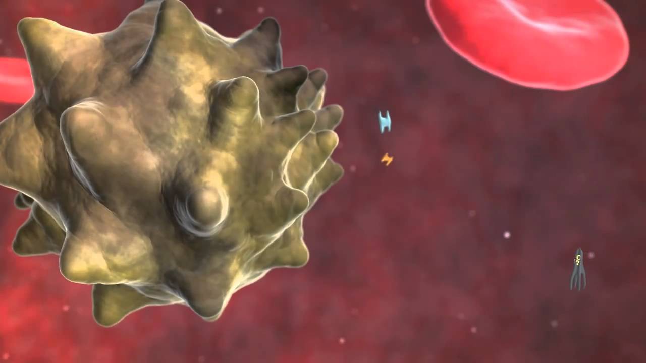
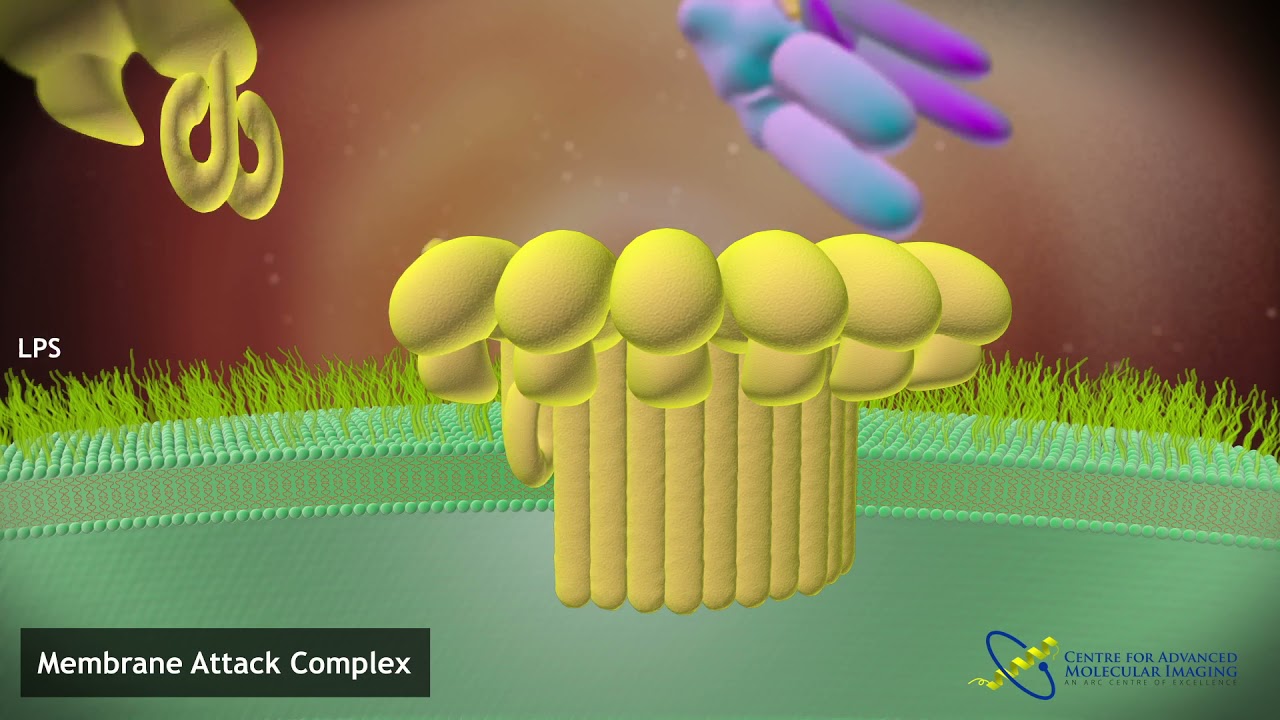

One of the best known host defense mechanisms of the complement system is its ability to split up many bacteria, enveloped virus and nucleated cells. This is mediated by a large macromolecular structure called the Membrane Attack Complex. MAC starts with C5b which associates with C6, 7 and 8.
The Membrane Attack Complex (MAC) is a crucial component of the immune system, specifically part of the complement system, which plays a vital role in innate immunity. The complement system is a group of proteins that work together to fight infections and clear pathogens from the body. The MAC is the terminal structure formed by the complement cascade, and its primary function is to lyse and eliminate pathogenic cells, such as bacteria and viruses, by creating pores in their membranes.
The Complement System: An Overview
The complement system consists of over 30 proteins that circulate in the blood and tissue fluids. These proteins are primarily produced by the liver and are present in an inactive form. Upon activation, they participate in a cascade of reactions that enhance the ability of antibodies and phagocytic cells to clear microbes and damaged cells, promote inflammation, and attack the pathogen's plasma membrane.
Pathways of Complement Activation
There are three main pathways through which the complement system can be activated:
- Classical Pathway: This pathway is initiated by the binding of the C1 complex to antibodies that are attached to antigens on the surface of pathogens.
- Lectin Pathway: This pathway is activated by the binding of mannose-binding lectin (MBL) to mannose residues on the surface of pathogens.
- Alternative Pathway: This pathway is continuously active at a low level and can be amplified in the presence of pathogens. It is initiated by the spontaneous hydrolysis of C3, a central component of the complement system.
All three pathways converge at the activation of C3, leading to the formation of C3 convertase, which cleaves C3 into C3a and C3b. The subsequent steps lead to the formation of C5 convertase, which cleaves C5 into C5a and C5b, initiating the assembly of the MAC.
Formation of the Membrane Attack Complex
The formation of the MAC involves a series of steps that result in the creation of a pore in the target cell's membrane:
- C5b Formation: The cleavage of C5 by C5 convertase results in the formation of C5b, which is the first component of the MAC.
- Assembly of C5b-6-7 Complex: C5b binds to C6 and C7, forming a complex that attaches to the target cell membrane.
- Insertion of C8: The C5b-6-7 complex recruits C8, which inserts into the lipid bilayer of the target cell membrane.
- Polymerization of C9: Multiple C9 molecules bind to the C5b-6-7-8 complex, forming a ring-like structure that creates a pore in the membrane.
The pore formed by the MAC disrupts the integrity of the target cell's membrane, leading to cell lysis and death. This process is particularly effective against Gram-negative bacteria, which have an outer membrane that is susceptible to MAC-mediated lysis.
Biological Significance of the MAC
The MAC plays a critical role in the immune defense against a variety of pathogens. Its ability to directly lyse cells makes it an essential component of the body's first line of defense. Some of the key functions and implications of the MAC include:
- Pathogen Elimination: By forming pores in the membranes of pathogens, the MAC directly contributes to their destruction and removal from the body.
- Immune Regulation: The complement system, including the MAC, helps regulate the immune response by promoting inflammation and recruiting immune cells to sites of infection.
- Protection Against Autoimmunity: Proper regulation of the complement system, including the MAC, is crucial to prevent damage to host tissues and avoid autoimmune diseases.
Clinical Implications and Disorders
While the MAC is an essential component of the immune system, its dysregulation can lead to various clinical conditions:
- Complement Deficiencies: Deficiencies in complement components, including those involved in MAC formation, can lead to increased susceptibility to infections, particularly with Neisseria species.
- Paroxysmal Nocturnal Hemoglobinuria (PNH): This rare disorder is characterized by the destruction of red blood cells due to uncontrolled complement activation and MAC formation on the surface of erythrocytes.
- Atypical Hemolytic Uremic Syndrome (aHUS): This condition involves excessive complement activation, leading to the formation of MAC on endothelial cells, resulting in cell damage and thrombotic microangiopathy.
The mystery behind membrane insertion: a review of the complement membrane attack complex
The membrane attack complex (MAC) is an important innate immune effector of the complement terminal pathway that forms cytotoxic pores on the surface of microbes. Despite many years of research, MAC structure and mechanism of action have remained elusive, relying heavily on modelling and inference from biochemical experiments. Recent advances in structural biology, specifically cryo-electron microscopy, have provided new insights into the molecular mechanism of MAC assembly. Its unique ‘split-washer’ shape, coupled with an irregular giant β-barrel architecture, enable an atypical mechanism of hole punching and represent a novel system for which to study pore formation. This review will introduce the complement terminal pathway that leads to formation of the MAC. Moreover, it will discuss how structures of the pore and component proteins underpin a mechanism for MAC function, modulation and inhibition.
The complement system, composed of over 35 proteins found in the plasma or bound to host cells, forms an integral part of the early immune response, Three major complement cascades, the classical, the alternative and the mannose-binding lectin pathways, can activate the terminal pathway, including the formation of the membrane attack complex (MAC).
MAC can form on and directly kill Gram-negative bacteria]. It is particularly important in combatting Neisseria meningitidis, with genetic deficiencies in MAC components leading to recurrent infections MAC pores can cause cell death by osmotic flux, and it has been postulated that the assembled pore may allow the passage of lysozymes across the outer membrane to degrade the peptidoglycan layer. Although the translocation of lysozyme through the MAC is implied through in vitro experiments, this concept is also supported by the known role of a close homologue, perforin. In the case of perforin, there is translocation of a range of proteins including granzymes from cytotoxic T cell granules into the cytoplasm of target cells to induce apoptosis.
While MAC function has been studied predominantly in the context of Gram-negative bacteria, it has also been shown to assemble on the surface of parasites [12], Gram-positive bacteria [13] and unwanted assembly on host cells. In nucleated host cells, membrane disruption results in cell death by apoptosis [14–16] or by lysis if sufficient MAC is present. Unlike erythrocytes, which are lysed by a single channel [18], nucleated cells can shed deposited pores in order to overcome the effects of the MAC In some circumstances, sublytic levels of MAC are found to be pro-survival, which may influence nearby cells during an inflammatory response . MAC deposition is implicated in a number of signal transduction pathways , such as G-protein and PI3 K signalling, and has been associated with platelet activation in host cells