Complement System
Return to Hematology Menu
Return to Immune System Menu
VIDEOS
Introduction
Animation
Animation
Chemicals of Inflammation - Plasma protein - Complement System, C3, C3a, C5, C5a
Kidney Disease
ABOUT
The complement system is composed of more than 30 heat-labile plasma proteins . Although complement activation plays a key role in innate immune defenses against invading bacteria, over-activation of complements leads to many inflammatory diseases including sepsis
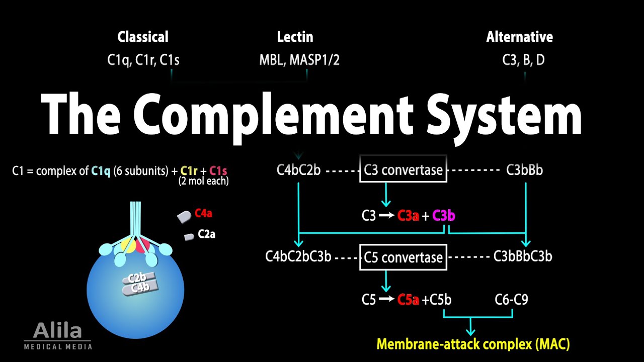
The complement system acts as an enzymatic cascade through a variety of protein-protein interactions, and complement activation occurs after a variety of different stimuli. Three well-known pathways are involved in complement activation: classical pathway, mannose-binding lectin (MBL) pathway, and alternative pathway.
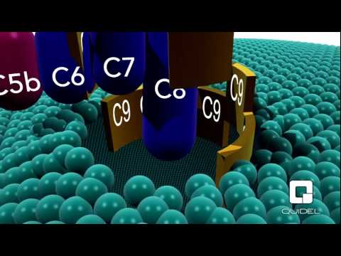
All the three pathways result in a series of enzymatic cleavage reactions, leading the formation of C3 convertase, at which the three pathways converge. C3 convertase can lead to the formation of C3a, C3b, C5a, C5b, C6, C7, C8, and C9, among which C5b, C6, C7, C8, and C9 form a membrane attack complex (C5b-9), which is used by host to lyse gram-negative bacteria.
The Coagulation pathway was recently suggested as a novel pathway of complement activation acting-independently of the formation of canonical C3/C5 convertases. In this pathway, thrombin functions as a C5 convertase in the absence of C3, leading to the production of C5a and formation of C5b-9.
Moreover, in multiple trauma patients, factor VII-activating protease (FASP), which was activated by circulating nucleosomes released from necrotic cells, interacted with complement proteins in plasma, and cleaved C3 and C5 to produce C3a and C5a. However, the mechanistic basis underlying the interaction between coagulation pathway and complement pathway remains poorly understood.
Complement as well as other parts of contact polysystem is based on stepwise limited proteolysis. In some kind, it is way of biological amplification of reactions because in the initial stage of complement activation, only a few molecules enter into reaction. On next step of the cascade, we have dozens of molecules, then hundreds of molecules, thousands of molecules. So, step by step, the overall effect is enhanced like in a snow avalanche. And, this is a way of biological amplification of reaction. It is characteristic for compliment system, for the coagulation system, for fibrinolysis, and to a certain degree for kinins also. The end product of this complement system is cytolytic. It can make a hole in the membrane of the target cell.
For example, self-cell in allergic reaction or bacterial cell in infectious inflammation. And in fact, the side products of this activation, cleavage peptides, are responsible for various effects regulating exudation, chemotaxis, switch of immunoglobulin classes and so on.
Typical pathways of complement activation.
Complement is a mediator of inflammation. The final product of this membrane attacking complex is responsible for lysis ofa target cell and the byproducts, anaphylatoxins, are side fragments of compliment, short peptides with known cytolytic bio-regulating action. For example, they increase exudation and vascular permeability is increased by them.
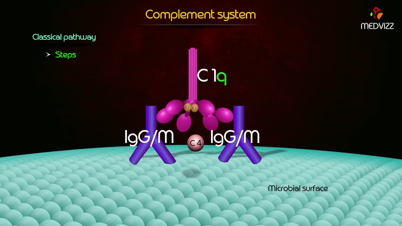
There are three pathways of activation for this system. The classic pathway, by the way, is realized if the complement is activated by antibodies in the course of immune response. And the alternative pathway, which can be activated directly without antibody participants by components of microorganisms, and also relatively, by a recently discovered lectin pathway or mannose-depending pathway, which is also activated outside of immunological interactions, even without antibodies. But generally speaking, compliment is a blind dog, and compliment needs some guide, which can indicate a target and the guide which indicates a target for that blind dog is an antibody.
Complement is assembled on the surface of a cell matched by antibodies.
Complement is responsible for complement-dependent cytotoxicity. It means the activation of complement system via the classical pathway is able to cause lysis of a cell on the surface of which the membrane attacking complex, C five, C nine is assembled. At the same time, fragments of complement factors arising from stepwise proteolysis are important mediators of accompanying inflammatory events. First of all, they are essential in exudation. As a result of this activation, the target cell dies and this complement depending cytotoxicity is broadly involved in immune and non-immune inflammation as a mechanism of secondary alteration.
Source: Lectures by Leonid P. Churilov, M.D, Ph.D, Chairman of Pathology Dept. at St. Petersburg State University
Glycine, serine and threonine metabolism confounds efficacy of complement-mediated killing Serum resistance is a poorly understood but common trait of some difficult-to-treat pathogenic strains of bacteria. Here, we report that glycine, serine and threonine catabolic pathway is down-regulated in serum-resistant Escherichia coli, whereas exogenous glycine reverts the serum resistance and effectively potentiates serum to eliminate clinically-relevant bacterial pathogens in vitro and in vivo. We find that exogenous glycine increases the formation of membrane attack complex on bacterial membrane through two previously unrecognized regulations: 1) glycine negatively and positively regulates metabolic flux to purine biosynthesis and Krebs cycle, respectively. 2) α-Ketoglutarate inhibits adenosine triphosphate synthase, which in together promote the formation of cAMP/CRP regulon to increase the expression of complement-binding proteins HtrE, NfrA, and YhcD. The results could lead to effective strategies for managing the infection with serum-resistant bacteria, an especially valuable approach for treating individuals with weak acquired immunity but a normal complement system.
FUNCTION IN GALT
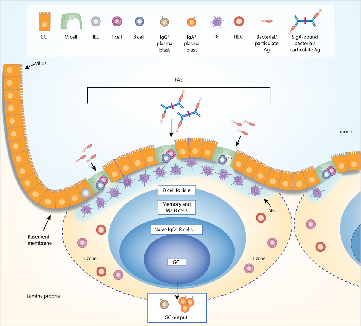
The complement system in the gut-associated lymphoid tissue (GALT) exhibits several unique adaptations tailored to the gut’s microenvironment, balancing pathogen defense with commensal microbiota tolerance. Here are the key features:
Localized Production and Microbiome Dependency
- Stromal cell synthesis: Gut complement component C3 is predominantly produced by stromal cells in intestinal lymphoid follicles (LFs), not just hepatocytes1. This local synthesis allows rapid, tissue-specific responses.
- Microbiota-driven regulation: Intestinal C3 levels are directly influenced by the composition of the gut microbiota15. For example, germ-free mice show reduced C3 levels, while colonization with specific bacteria restores it.
Luminal Secretion and Pathogen Clearance
- Active secretion into the gut lumen: C3 is secreted into the intestinal lumen, where it mediates opsonization and phagocytosis of pathogens like Citrobacter rodentium and enterohemorrhagic E. coli (EHEC)1.
- Enhanced defense during infection: Luminal C3 levels spike during enteric infections, directly correlating with pathogen clearance1. C3-deficient mice exhibit increased epithelial invasion by pathogens, underscoring its protective role.
Synergy with Mucosal Antibodies
- Interaction with IgT/IgZ in teleosts: In fish, IgT+ B-cells release antibodies that activate complement-mediated lysis or coat microbes to block translocation4.
- Mammalian parallels: While mammals lack IgT, IgA likely plays a similar role in coating pathogens and recruiting complement for neutralization.
Structural and Functional Adaptations
- Diffuse lymphoid organization: Unlike systemic lymphoid tissues, GALT in many species (e.g., teleost fish) lacks organized structures like Peyer’s patches, relying instead on diffusely distributed leukocytes (e.g., intraepithelial lymphocytes) to interact with complement47.
- Chemotaxis over lysis: The complement system in GALT prioritizes chemotaxis (via C5a, C3a) to recruit macrophages and neutrophils, rather than direct cell lysis, minimizing collateral damage to the epithelial barrier36.
Immune Tolerance and Homeostasis
- Balancing act: GALT’s complement activity is tightly regulated to avoid attacking commensal bacteria. Microbiota-derived signals modulate C3 production, ensuring responses are pathogen-specific18.
- Epithelial collaboration: Gut epithelial cells contribute to complement protein synthesis (e.g., via Paneth cells), creating a frontline defense layer65.
Evolutionary Conservation
- Cross-species relevance: Similar mechanisms exist in marsupials and fish, highlighting the evolutionary conservation of gut-specific complement functions. For example, stromal cell-driven C3 production and microbiota interactions are observed in both mice and humans14.
In summary, the gut complement system is uniquely compartmentalized, microbiome-dependent, and tailored to neutralize pathogens while preserving epithelial integrity and commensal harmony. Its integration with mucosal antibodies and stromal cell activity underscores its specialized role in intestinal immunity.
HOW DOES THE GUT MICROBIOTA INFLUENCE THE COMPLEMENT SYSTEM IN THE GUT
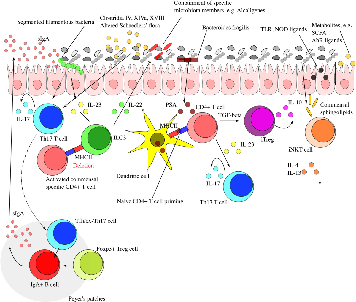

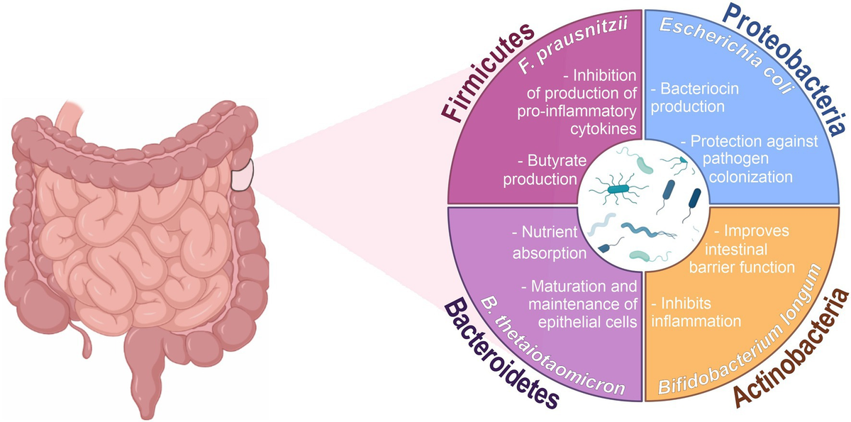
The gut microbiota plays a central role in regulating the complement system within the intestinal environment, primarily through microbiota-dependent production of complement components and fine-tuned immune responses. Here are the key mechanisms:
Microbiota-Driven C3 Production
- Local synthesis by stromal cells: Complement component C3—central to complement activation—is predominantly produced by stromal cells in intestinal lymphoid follicles rather than hepatocytes (the primary source in systemic circulation). This localized production is directly regulated by the composition of the gut microbiota14.
- Host-specific modulation: Germ-free mice exhibit significantly reduced intestinal C3 levels, which are restored upon colonization with specific commensal bacteria1. In humans, variations in microbiota composition correlate with individual differences in luminal C3 levels, influencing susceptibility to enteric infections14.
Pathogen Clearance Without Commensal Harm
- Luminal secretion for defense: C3 is actively secreted into the gut lumen, where it opsonizes pathogens (e.g., Citrobacter rodentium, EHEC) to enhance phagocytosis by immune cells14. This mechanism is critical during infections, as C3-deficient mice show increased epithelial invasion by pathogens1.
- Avoidance of commensal lysis: The gut complement system lacks membrane attack complex (MAC) components, preventing lytic damage to commensal bacteria despite C3 deposition on microbial surfaces4.
Microbiota-Complement Crosstalk in Homeostasis
- Balanced immune activation: Commensal microbes suppress excessive complement activation to maintain tolerance. For example, microbiota-derived signals regulate C3 production to ensure pathogen-specific responses while sparing beneficial bacteria42.
- Epithelial collaboration: Gut epithelial cells and stromal cells work synergistically to adjust complement protein levels in response to microbial shifts, creating a dynamic defense layer14.
Implications for Disease
- Dysbiosis-linked vulnerabilities: Altered microbiota composition (dysbiosis) reduces luminal C3 levels, increasing susceptibility to diarrheal infections1. This is particularly relevant in conditions like obesity and malnutrition, where dysbiosis is common1.
- Therapeutic potential: Modulating gut microbiota to enhance C3 production could offer strategies to combat enteric pathogens or mitigate chronic inflammation14.
In summary, the gut microbiota acts as a master regulator of the intestinal complement system, tailoring immune responses to neutralize pathogens while preserving commensal harmony. This relationship underscores the importance of microbial balance in maintaining gut immune efficacy.
HOW DOES THE COMPLEMENT SYSTEM HELP PROTECT AGAINST ENTERIC INFECTIONS IN THE GUT?
The complement system plays a critical role in protecting against enteric infections through gut-specific adaptations that enhance pathogen clearance while maintaining mucosal integrity. Key mechanisms include:
Localized C3 Production by Stromal Cells
- Intestinal lymphoid follicle (LF) stromal cells are the primary source of gut-specific complement component C3, rather than hepatocytes (which produce systemic complement)12.
- This localized synthesis enables rapid, microbiota-dependent responses. Germ-free mice exhibit reduced intestinal C3 levels, which normalize after microbial colonization, highlighting the microbiome's role in regulating C3 production12.
Luminal C3 Secretion and Opsonophagocytosis
- During infections (e.g., Citrobacter rodentium, enterohemorrhagic E. coli), C3 is actively secreted into the gut lumen, where it opsonizes pathogens for phagocytosis by immune cells12.
- C3-deficient mice show increased epithelial invasion by pathogens, demonstrating its necessity for containment. In wild-type mice, C3-mediated phagocytosis effectively clears pathogens like C. rodentium12.
Synergy with Mucosal Immune Defenses
- IgA collaboration: While not directly producing lytic damage, C3 works alongside secretory IgA to neutralize pathogens. IgA coats microbes to block epithelial translocation, while C3 enhances phagocytic clearance3.
- Epithelial barrier reinforcement: Complement activation recruits immune cells (via C5a/C3a chemotaxis) to infection sites, strengthening mucosal defenses without disrupting commensal microbiota13.
Microbiota-Driven Immune Balance
- Commensal microbes regulate C3 levels to maintain a pathogen-specific response. Dysbiosis reduces luminal C3, increasing susceptibility to diarrheal infections12.
- In humans, individual variations in microbiota composition correlate with intestinal C3 levels, influencing infection resistance12.
Avoidance of Commensal Damage
- The gut complement system prioritizes opsonization over membrane attack complex (MAC) formation, preventing lytic damage to beneficial bacteria despite C3 deposition on microbial surfaces13.
In summary, the gut complement system protects against enteric pathogens through microbiota-regulated C3 production, luminal opsonophagocytosis, and collaboration with mucosal defenses—strategies that balance pathogen clearance with commensal preservation123.
ACTIVATORS OF COMPLEMENT SYSTEM
Almond Mushroom
Almond mushroom helps fight off fungal infections by activating the complement system. Phagocytosis of articulate activators of the human alternative complement pathway through monocyte beta-glucan receptors
Activation of the alternative complement pathway by Agaricus blazei Murill Components of Agaricus blazei Murill have been demonstrated to have a wide range of immunopotentiating activities. The present study was designed to evaluate the effect of A. blazei Murill upon activation of the complement system in human serum in vitro. Additional studies were performed to determine the cytotoxic effect of complement-opsonized particles of A. blazei Murill against human tumor cells in culture. A fine particle of A. blazei Murill (ABP), prepared by mechanical disruption, was used throughout the experiments. ABP activated the human complement system via the alternative pathway in human serum. Activation of the alternative pathway was both time- and dose-dependent. When the particles from fruiting bodies of A. blazei Murill (ABP-F) were reacted with human serum, the formation of complement-opsonized ABP, iC3b-ABP-F complexes, and binding of the complexes to human peripheral blood monocytes, were demonstrated in vitro by immunofluorescence. Further, the resident human peripheral nucleated cells incubated in the presence of iC3b-ABP-F complexes inhibited the proliferation of human tumor cell line TPC-1 in vitro.
IgM (Immunoglobulin M): The first antibody produced in response to an infection, IgM is effective in forming complexes with antigens and activating the complement system.
- It can stimulate the complement system leading to immune hyper-reactivity.
Sambucus ebulus fruits modulate inflammation and complement system activity in humans.