Crystallography
In 1784 René-Just Haüy* showed that crystals with the same composition will have the same internal nucleus, even if their external shapes are different and that crystal forms are based on fixed laws.
The law of symmetry was further defined by Hessel. In 1830 he declared that "geometric constraints limit the number of crystal classes to exactly 32, and that only twofold, threefold, fourfold and sixfold axes of rotational symmetry are possible in minerals."
At the end of the nineteenth century Roentgen* discovered X-rays and set the stage for studying crystals at the molecular and even atomic scale.
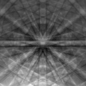
In 1911, Von Laue* passed a narrow X-ray through a copper sulphate crystal; the resulting pictures on the photographic plate could only be explained by a regular arrangement of atoms with spacings comparable to the length of X-rays.
The Nobel Prize in Physics was awarded to a father-and-son team ( Bragg*) a few years later using Von Laue’s X-ray diffraction technique to calculate the exact position of atoms in crystal structures.
The science of crystallography has continued to be explored, as instruments capable of ever more precise and miniscule examination are developed.
ONELINER: Crystallography is experimental science of determining the arrangement of atoms in crystalline solids
OTHER CONNECTIONS: None
BIBLIOGRAPHY: http://www.newadvent.org/cathen/07152a.htm;
http://geology.csupomona.edu/drjessey/class/gsc215/minnotes1.htm
Biology[[]
X-ray crystallography is the primary method for determining the molecular conformations of biological macromolecules, particularly protein and nucleic acids such as DNA and RNA. In fact, the double-helical structure of DNA was deduced from crystallographic data. The first crystal structure of a macromolecule was solved in 1958, a three-dimensional model of the myoglobin molecule obtained by X-ray analysis.[6] The Protein Data Bank (PDB) is a freely accessible repository for the structures of proteins and other biological macromolecules. Computer programs such as RasMol, Pymol or VMD can be used to visualize biological molecular structures.
Neutron crystallography is often used to help refine structures obtained by X-ray methods or to solve a specific bond; the methods are often viewed as complementary, as X-rays are sensitive to electron positions and scatter most strongly off heavy atoms, while neutrons are sensitive to nucleus positions and scatter strongly even off many light isotopes, including hydrogen and deuterium. Electron crystallography has been used to determine some protein structures, most notably membrane proteins and viral capsids.
Contribution of women to X-ray crystallography[[]
A number of women were pioneers in X-ray crystallography at a time when they were excluded from most other branches of physical science.[7]
Kathleen Lonsdale was a research student of William Henry Bragg, who with his son Lawrence founded the science of X-ray crystallography at the beginning of the 20th century. She is known for both her experimental and theoretical work. Bragg had 11 women research students out of a total of 18. Kathleen joined his crystallography research team at the Royal Institution in London in 1923, and after getting married and having children, went back to work with Bragg as a researcher. She confirmed the structure of the benzene ring, carried out studies of diamond, was one of the first two women to be elected to the Royal Society in 1945, and in 1949 was appointed the first female tenured professor of chemistry and head of the Department of crystallography at University College London.[8]
Kathleen always advocated greater participation of women in science and said in 1970: "Any country that wants to make full use of all its potential scientists and technologists could do so, but it must not expect to get the women quite so simply as it gets the men. ... It is utopian, then, to suggest that any country that really wants married women to return to a scientific career, when her children no longer need her physical presence, should make special arrangements to encourage her to do so?".[9] During this period, Kathleen began a collaboration with William T. Astbury on a set of 230 space group tables which was published in 1924 and became an essential tool for crystallographers.
[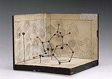
Molecular model of penicillin by Dorothy Hodgkin, 1945
In 1932 Dorothy Hodgkin joined the laboratory of the physicist John Desmond Bernal, who was a former student of Bragg, in Cambridge, UK. She and Bernal took the first X-ray photographs of crystalline proteins. Hodgkin also played a role in the foundation of the International Union of Crystallography. She was awarded the Nobel Prize in Chemistry in 1964 for her work using X-ray techniques to study the structures of penicillin, insulin and vitamin B12. Her work on penicillin began in 1942 during the war and on vitamin B12 in 1948. While her group slowly grew, their predominant focus was on the X-ray analysis of natural products. She is the only British woman ever to have won a Nobel Prize in a science subject.

Photograph of DNA (photo 51), Rosalind Franklin, 1952
Rosalind Franklin took the X-ray photograph of a DNA fibre that proved key to James Watson and Francis Crick's discovery of the double helix, for which they both won the Nobel Prize for Physiology or Medicine in 1962. Watson revealed in his autobiographic account of the discovery of the structure of DNA, The Double Helix,[10] that he had used Rosalind's X-ray photograph without her permission. Franklin died of cancer in her 30s, before Watson received the Nobel Prize. Franklin also carried out important structural studies of carbon in coal and graphite, and of plant and animal viruses.
Isabella Karle of the United States Naval Research Laboratory developed an experimental approach to the mathematical theory of crystallography. Her work improved the speed and accuracy of chemical and biomedical analysis. Yet only her husband Jerome shared the 1985 Nobel Prize in Chemistry with Herbert Hauptman, "for outstanding achievements in the development of direct methods for the determination of crystal structures". Other prize-giving bodies have showered Isabella with awards in her own right.
Women have written many textbooks and research papers in the field of X-ray crystallography. For many years Lonsdale edited the International Tables for Crystallography, which provide information on crystal lattices, symmetry, and space groups, as well as mathematical, physical and chemical data on structures. Olga Kennard of the University of Cambridge, founded and ran the Cambridge Crystallographic Data Centre, an internationally recognized source of structural data on small molecules, from 1965 until 1997.
Jenny Pickworth Glusker, a British scientist, co-authored Crystal Structure Analysis: A Primer,[11] first published in 1971 and as of 2010 in its third edition. Eleanor Dodson, an Australian-born biologist, who began as Dorothy Hodgkin's technician, was the main instigator behind CCP4, the collaborative computing project that currently shares more than 250 software tools with protein crystallographers worldwide.