RNP Structure of CoV
Overview to N-RNA packaging
(Ross 2022 - PMID: 35131265) Multiple studies suggest that a single strand of SARS-CoV-2 gRNA forms dense, locally ordered ribonucleoprotein (RNP) regions consisting predom- inantly of N protein associated with the gRNA strand (3–5). Locally ordered RNPs may be further organized into more complex arrangements via clustering of RNPs in particular stoichiometries and geometries (3, 4), although other evidence and prior models of the SARS-CoV N protein suggest a more linear, helical RNP arrangement (2, 5, 6). These RNPs preferentially accumulate on curved membranes (3), indicating either that RNP association aids in membrane curvature dur- ing nascent virion formation, or that curved membranes are the preferred recruitment surface for RNPs. The N protein also interacts with a luminal domain (i.e., in the interior of virions) of the M protein, which was proposed as a possible mechanism for mediating recruitment of N-containing RNPs to the ERGIC membrane (7). Some evidence suggests that the N protein of both SARS-CoV and SARS-CoV-2 may also interact with the E protein (8, 9). While precise detail regarding the interactions and arrangements of individual molecules within intact virions is still forthcoming, the N protein clearly plays a central role in gRNA compaction and organization in SARS-CoV-2 virions.
EM of RNP
(2009 pmid: 19124777) Cryo EM Tomography
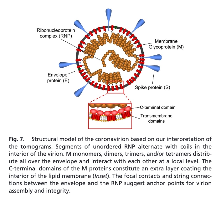
- Focal pairs revealed the existence of an extra internallayer, - formed by the C-terminal domains of M.
- interior - coiled structures and tubular shapes - helical nucleocapsid model.
- no evidence of a shelled core. ribonucleoprotein seems to be exten-sively folded onto itself, assuming a compact structure that tends to closely follow the envelope at a distance of 4 nm.
- Focal contactpoints and thread-like densities connecting the envelope and the ribonucleoprotein
(2017 pmid: 28044277)
- A dense interior core corresponding to the intertwined RNP is encapsulated inside the envelope
- RNP extracted from the virus - negative staining electron microscopy - coronavirus RNP might be a long helix with a diameterbetween 9 nm to 16 nm (Macneughton and Davies,1978).
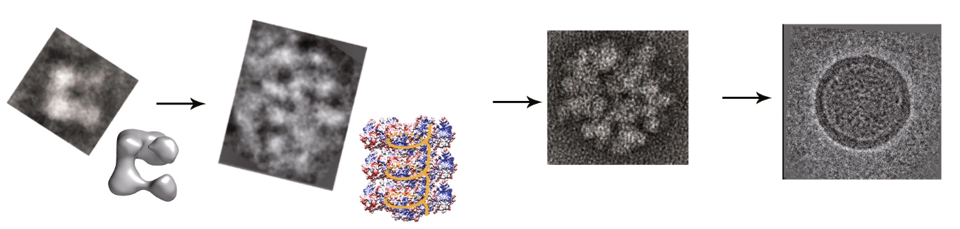
- ∼3% CHAPS. RNPs were released after the treatment. The released RNPs are in either a loose filament structure or in a compact flower-like assembly that may be similar to the intact RNP assembly in virus particles. MHV RNP - Cryo and EM - RNPs. - also found that the isolated intact RNPs degraded into small pothook-like subunits. These small subunits could be thebuilding blocks of the long loose helical and the supercoiled flower-like RNP structures.
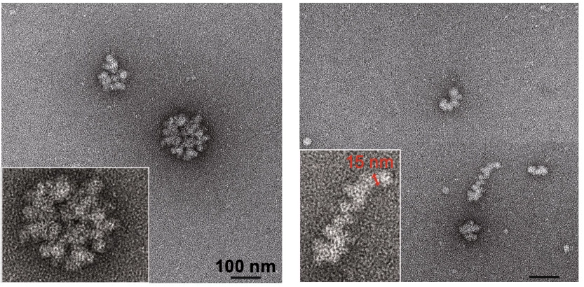
- fitted N into 3D map - consistent with SAX (Chang et al.,2009); crystal packing of CTD as octamer (Chang et al.,2014); In the model weproposed, two subunits pack head to head into a helicalfilament. The width of the modeledfilament is twice of thesubunit (∼15 nm). Distance between the repeating strands,which is the distance of the two arms of the pothook, isaround 7 nm. modeling... lots of assumptions
RNA structure of the CoV2 genome - SHAPE
see: RNA Genome Structure