Nucleocapsid Protein (NP)
Intro
(Lu, Corbett NatCom 2021 PMID: 33479198) The RNA-binding N protein plays two major roles in the coronavirus life cycle.
Its primary role is to assemble with genomic RNA into the viral RNA–protein (vRNP) complex, and mediate vRNP packaging into virions via poorly understood interactions between the N and M proteins8–10.
Second, the N protein localizes to RTCs at early stages of infection, where it is thought to facilitate viral RNA synthesis and translation by recruiting host factors and promoting RNA template switching11–15.
(Lu, Corbett NatCom 2021 PMID: 33479198)
N proteins self-associate into dimers, tetramers, and larger oligomers that are thought to form the basis for assembly of the vRNP complex21,27–29.
In the virion, the N protein mediates assembly of a helical filament-like vRNP complex3,30–32. Cryo-electron tomography (cryo-ET) of SARS-CoV-2 virions has revealed a more granular vRNP structure, with each virion containing 35–40 individual vRNP complexes that each adopt a cylindrical shelllike structure ~15 nm in diameter4,33.
Within the virion, membrane-proximal vRNPs show a characteristic orientation with respect to the membrane, suggesting that they specifically interact with the membrane-associated M protein33. Within the vRNP, tentative modeling based on known NTD and CTD structures suggests a specific assembly with ~800 nt of genomic RNA (30 kb ÷ ~38 vRNPs) wrapped around ~12 copies of the N protein33. Individual vRNPs could also form linear stacks resembling helical filaments4,33, reconciling the apparent conflict with earlier observations and suggesting that betacoronavirus vRNPs likely adopt a broadly conserved architecture.
PMID: 35874957 - 2022
N protein exhibits three important roles in coronavirus life cycle
- the primary role is to assemble with genomic RNA into the viral RNA-protein (vRNP) complex ^22. Virion formation occurs via the accumulation of the SARS-CoV-2 structural proteins [S, E, M and N] and gRNA at the ER-Golgi intermediate compartment (ERGIC) membrane. The N protein can interact with a luminal domain of the M protein through C-terminal dimerization domain (CTD) ^(247-365), which may be essential for mediating the recruitment of N-containing RNPs to the ERGIC membrane ^23. Previous study also suggests that the interaction between N protein and the E protein may plays an important role in SARS-CoV-2 assembly ^24. Together with N protein, a single strand of SARS-CoV-2 gRNA forms dense, locally ordered ribonucleoprotein (RNP) regions which may be further organized into more complex arrangements ^25-27.
- liquid-liquid phase separation (LLPS) with viral genome RNA and potentially facilitates the viral assembly. Many RNA-binding proteins have been found to undergo LLPS with RNA to participate the biological and disease processes ^28-32. The LLPS is dependent on the length and concentration of ssRNA. What is more, N protein forms typical sphere-like droplets with short ssRNAs, but not solid-like structures with long ssRNAs. The free Zn2+ in cytosol is essential for N protein/RNA LLPS ^21. It is also reported that viral RNA can induce assembly of the N protein into phase-separated condensates in vitro and pinpoints a ~40 residue region in the central intrinsically disordered region (IDR) with a key role in RNA-driven phase separation ^23.
- localize to replication transcription complexes (RTCs) at the early stages of infection, where it enhances replication and the transcription of viral RNA by recruiting host factors 15,33-36. Previous studies have demonstrated that N protein and RNA recruited components of the RNA polymerase (RdRp) complex (Nsp7, Nsp8 and Nsp12) are responsible for replicating viral gRNA 37,38. The phosphorylation of the SR domain of the N protein can decrease the degree of recruitment of RdRp components to condensates ^37. The interaction between N and Nsp3 is essential for viral replication. The N-terminal of N has an RNA-binding domain (N-NTD) that can bind the RNA genome, whereas, the C-terminal domain (N-CTD) can interact with the viral Nsp3 ^39.
Review (2014 pmid: 24418573)
(2020 pmid: 32363136)
- multifunctional RNA-binding protein necessary for viral RNA transcription and replication.
- forming helical ribonucleoproteins during the packaging of the viral RNA genome 10,11
- binding a leader RNA 8,12
- regulating viral RNA synthesis in replication/transcription, and modulating infected cell metabolism7-9.
- regulated host-pathogen interactions, such as actin reorganization, host cell cycle progression, and apoptosis13-15.
- highly immunogenic and abundantly expressed protein during infection, capable of inducing protective immune 16-19.
- Nucleocapsid (N) protein is translated from the 3′ end structural ORF35-37.
- sequence identities between SARS-CoV-2 with SARS-CoV, MERS-CoV, and HCoV-OC43 were 89.74%, 48.59%, 35.62%, respectively
(Giedroc 2009 pmid: 19782089)
- N has been shown to significantly enhance the efficiency of RNA replication.17
- important accessory factor in discontinuous transcription.18
- These multifunctional properties of CoV N are analogous to HIV-1 nucleocapsid protein19 and make N an attractive antiviral target.
- (Liu NAR 2006 pmid: 16971454) RNA binding is essential for infectivity
- (pmid: 28720894) the only structural protein that associates with RTCs9–11; Replicase-Transcriptase Complex
- NP recognices the PS Recognition of the PS
- (Peng 2020 Embo pmid: 32914439) to function as a viral suppressor of RNA silencing (VSR) through its double-stranded RNA-binding activity to combat host RNAi-mediated antiviral responses (Mu et al, 2020).
- (doi: 10.1101/2020.12.26.424450) N can constitute up to ~1% of expressed proteins during infection
Signaling functions of NP - NP - Signaling functions
Nucleocapsid synthesis in the life cycle of the virus
(Carlson MolCell 2020 PMID: 33248025) Using genomic (+) RNA as a template, the viral polymerase produces (-) RNA transcripts of subgenomic regions encoding the viral structural proteins (S, E, M, and N). Sub-genomic transcription involves a template-switching mechanismin which the polymerase completes transcription of a structural protein gene and then skips to a transcription-regulating sequence (TRS) at the 5’end of the genome, resulting in subge-nomic (minus) RNA fragments—which are then transcribed to produce (+) RNA for translation. N protein is encoded by the most abundant subgenomic RNA and is translated at high levels early in infection. The N protein is the most abundant viral protein inthe infected cell and accumulates in dynamic clusters at RTCs where it is thought to help promote the RNA structural rearrangements required for subgenomic transcription
Protein Data
P0DTC9 · NCAP_SARS2 UniProt
Sequence : MSDNGPQNQRNAPRITFGGPSDSTGSNQNGERSGARSKQRRPQGLPNNTASWFTALTQHG
KEDLKFPRGQGVPINTNSSPDDQIGYYRRATRRIRGGDGKMKDLSPRWYFYYLGTGPEAG
LPYGANKDGIIWVATEGALNTPKDHIGTRNPANNAAIVLQLPQGTTLPKGFYAEGSRGGS
QASSRSSSRSRNSSRNSTPGSSRGTSPARMAGNGGDAALALLLLDRLNQLESKMSGKGQQ
QQGQTVTKKSAAEASKKPRQKRTATKAYNVTQAFGRRGPEQTQGNFGDQELIRQGTDYKH
WPQIAQFAPSASAFFGMSRIGMEVTPSGTWLTYTGAIKLDDKDPNFKDQVILLNKHIDAY
KTFPPTEPKKDKKKKADETQALPQRQKKQQTVTLLPAADLDDFSKQLQQSMSSADSTQA
can be bought: Sino Biological
Domain architecture
(Peng 2020 Embo pmid: 32914439) Coronavirus N proteins have two conserved and independently folded structural domains, called the N-terminal domain (NTD) and C-terminal domain (CTD) (Chang et al, 2014). The two domains are connected by an intrinsically disordered region (IDR) called the central linker region (LKR). The LKR includes a Ser/Arg (SR)-rich region that contains putative phosphorylation sites (Peng et al, 2008). In addition, the N-NTD and N-CTD of coronaviruses are usually flanked by two IDRs, which are called N-arm and C-tail. Previous studies have revealed that N-NTD is responsible for RNA binding, NCTD for both RNA binding and dimerization, and the IDR for modulating the RNA-binding activity of N-NTD and N-CTD and oligomerization (Chang et al, 2014).
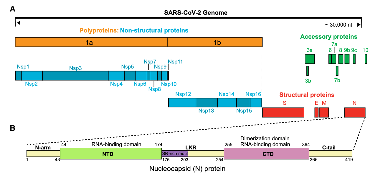
(Kuo Masters 2014 J Virol PMID: 24501403) Adjacent to the RNA-binding domains are intrinsically disordered segments (29) falling at the amino terminus (domain N1a), the carboxy terminus (spacer B and domain N3), and the center (domain N2a) of the N molecule. No functional role has been ascribed to domain N1a. Domain N3 has been shown to interact with the carboxy terminus of the M protein (30–33), and it also appears to participate in one type of N-N interaction (16). Domain N2a contains a serine- and arginine-rich region (Fig. 1A) that interacts with the replicase subunit nonstructural protein 3 (nsp3) to mediate a crucial early step in infection (17, 18, 34).
Overview to domain functions
(Lu, Corbett NatCom 2021 PMID: 33479198) The N-terminal domain (NTD) is thought to mediate a specific interaction with the viral genome’s packaging signal, and the C-terminal domain (CTD) forms a compact dimer that has been proposed to aid vRNP assembly16–21. These two domains are separated by a conserved central IDR containing a serine/arginine-rich region (SR) that is highly phosphorylated in infected cells22–26, and are flanked by less well-conserved IDRs at the N- and C-terminus. The C-terminal IDR of the related SARS-CoV N protein has been implicated in M protein binding, suggesting an important role for this domain in viral packaging9,10.
Functions of the RBD - NP - RNA binding domain (RBD)
Functions of the IDR - NP - RNA phase separation
RNA binding
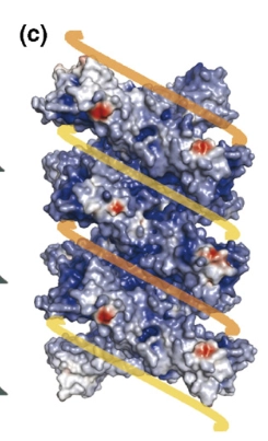
(pmid: 17379242) Nucleocapsids contain a large number of copies of the structural protein(s), which often polymerize through a selfassembly mechanism. Some viruses form helical capsids.
For details: NP-RNA interaction
-- SARS-CoV N protein can bind to DNA in vitro
-- N protein fragments containing the dimerization domain (residues 236–384) could also bind to an RNA packaging signal.17
(pmid: 28720894) cytoplasmic N molecules of MHV-infected cells constitutively assemble into oligomers through a process that does not require binding to genomic RNA.
-- model: constitutive N protein oligomerization allows the optimal loading of the genomic viral RNA into a ribonucleoprotein complex via the presentation of multiple viral RNA binding motifs.
Recognition of the Packaging signal
RBD or CTD ? XXXXXX (Lu, Corbett NatCom 2021 PMID: 33479198) vs others that say CTD XXX check
see: PS of Betacoronavirus lineage B (ie SARS-CoV)
NP dimerisation - Oligomerisation
see: NP - dimerisation domain
(Carlson MolCell 2020 PMID: 33248025) Under some conditions, the dimer self-associates to form oligomers that depend on multiple protein regions (Chang et al., 2013; Chen et al., 2007; Cong et al., 2017; He et al., 2004; Hurst et al., 2009; Luo et al., 2006; Luo et al., 2005; Ye et al., 2020; Zuwala et al., 2015). The biochemical features and function of these oligomers are not clear.
Inhibitiors for NP
(Peng 2020 Embo pmid: 32914439) small-molecule drugs can be developed by targeting the RNAbinding activity and the oligomerization of the N protein (Lin et al, 2014; Chang et al, 2016; Zhang et al, 2020b).
see: Nucleocapsid Project
Vaccine Targets for CoV-2
(Ahmed McCay 2020 Viruses pmid: 32106567) -- identified a set of B cell and T cell epitopes derived from the spike (S) and nucleocapsid (N) proteins