NP - RNA cryo structures
NP RNA binding domains - Cryo
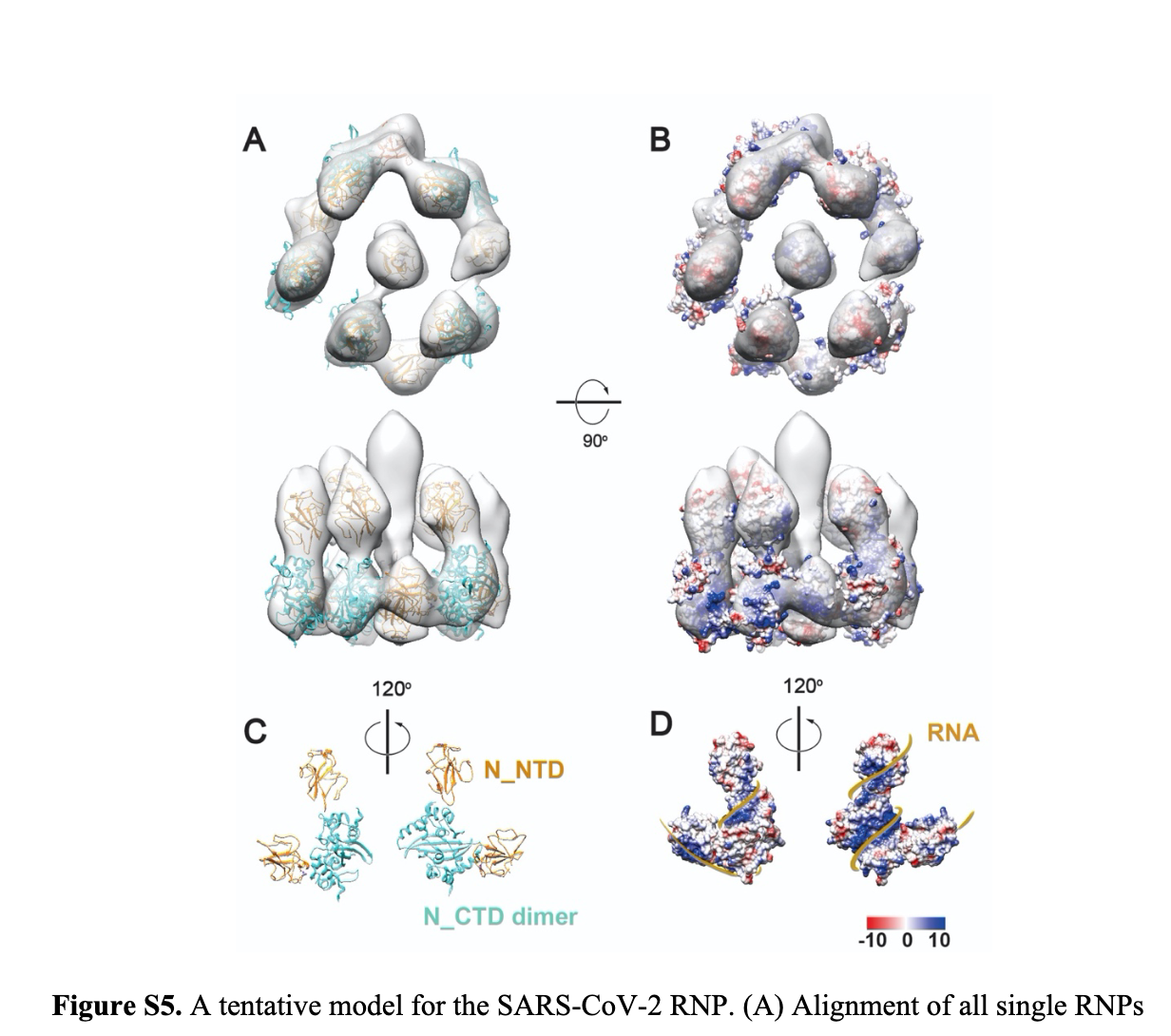
(pmid: 32766587) Recent high-resolution cryo-electron tomography of intact SARS-CoV-2 virions - individual viral RNA-protein complexes (vRNPs) adopt a characteristic shell-like architecture ∼15 nm in diameter, comprising ∼12 N proteins and ∼800 nt of RNA (4, (Neufeldt 2020 doi: 10.1101/2020.06.23.167064) ).
Negative-stain electron microscopy of soluble N+RNA complexes has revealed similar particles (52), suggesting that viral RNA packaging is mediated first by assembly of individual vRNPs along the genomic RNA, followed by condensation of these RNPs and recruitment to developing virions through interactions with the M protein at the cytoplasmic side of the ER-Golgi intermediate compartment (Fig. 6B) (4, 61).
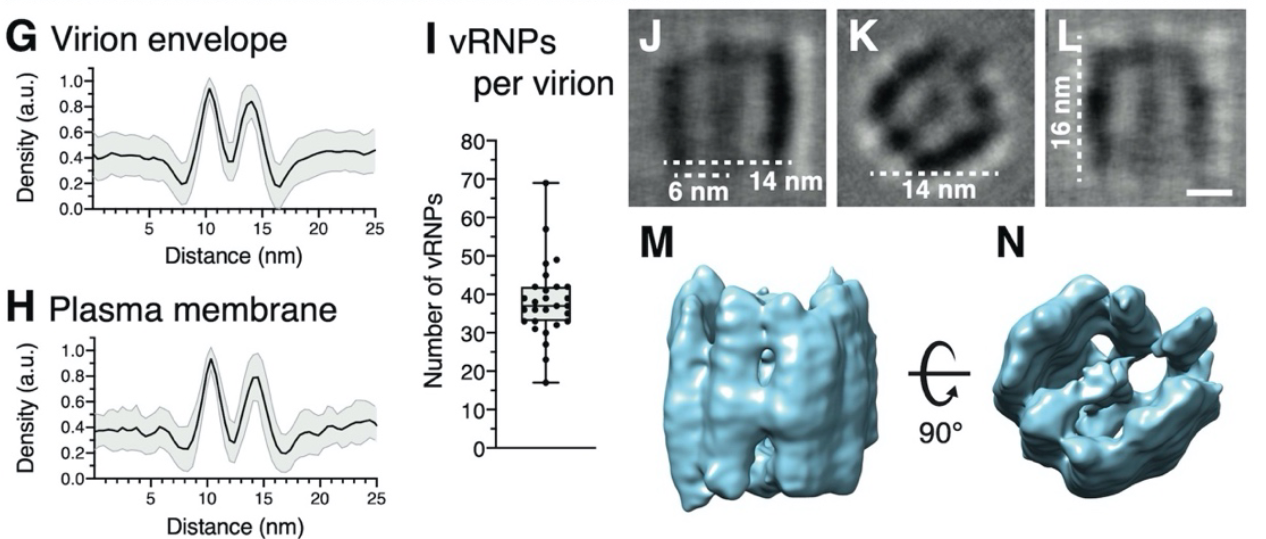
(Klein Chlanda 2020 doi: 10.1101/2020.06.23.167064) vRNPs associated with viral envelopes were often aligned in stacks, forming filaments with a width of approximately 14 nm.
- vRNPs yielded a compact, cylindrical assembly of 16 nm in length and a quasi-circular base with an approximate diameter of 14 nm
- multiple linearly aligned N proteins. Pillar-shaped densities form two densely packed curved walls opposing each other and surrounding a central density, separated from the walls by 6 nm.
- We propose that vRNPs are separate complexes organized like ‘beads on a string’ that allows for efficient packing of the unusually large vRNA genome into the virus particles while maintaining high sterical flexibility between the individual vRNPs required during their incorporation into budding virions.
(doi: 10.2139/ssrn.3654623)
classification of the RNPs revealed three classes: 1) closely packed against the envelope, 2) hexagonally and 3) triangularly packed RNPs (Figure4A).
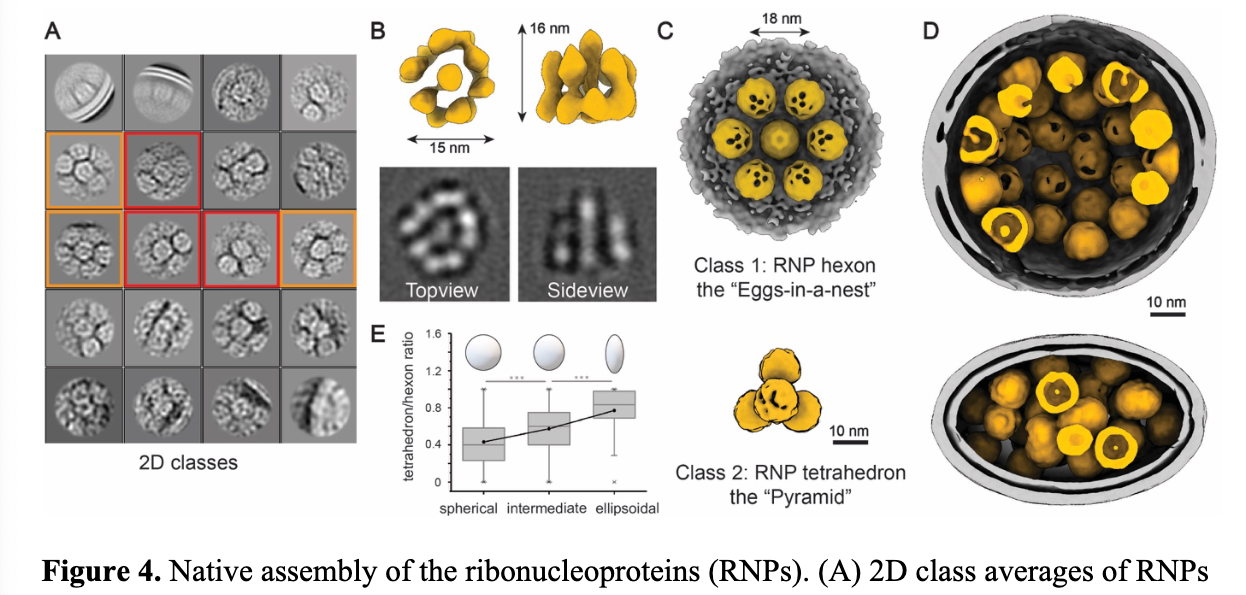
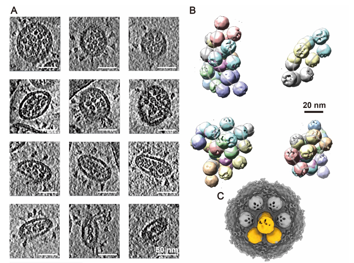
No virus with strictly ordered RNPs throughout the lumen was found by projections. We conclude that the native RNPs are highly heterogeneous, densely packed yet locally ordered in the virus, possibly interacting with the RNA in a beads-on-a-string stoichiometry.
RNA double stranded; measure number of particles and RNA length...
Octameric structure
pmid: 35551296 / 2021
CoV2 Ribonucleosome
PMID: 35664996 / 2022 Carlson/Morgan
each virion contains 35-40 viral ribonucleoprotein (vRNP) complexes, or ribonucleosomes, arrayed along the genome.
N-RNA: forms cylindrical 15-nm particles similar to the vRNP structures observed within coronavirus virions.
form in the presence of stem-loop-containing RNA and depend on regions of N protein that promote protein-RNA and protein-protein interactions.
Phosphorylation of N protein in its disordered serine/arginine (SR) region weakens these interactions and disrupts vRNP assembly. We propose that unmodified N binds stem-loop-rich regions in genomic RNA to form compact vRNP complexes within the nucleocapsid, while phosphorylated N maintains uncompacted viral RNA to promote the protein's transcriptional function.
NP-RNA binding dynamics
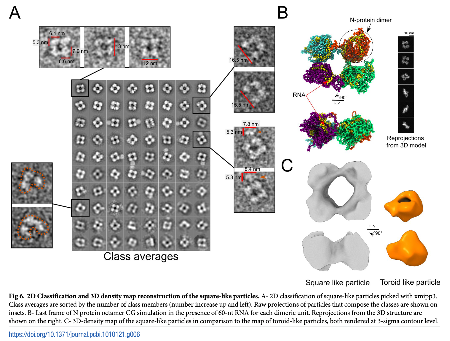
PMID: 35551296 / 2022 / Ribeiro/Oliviera : using experimental data from electron microscopy and biochemical/biophysical techniques combined with molecular modeling and molecular dynamics simulations
in the absence of RNA, the N protein formed structurally dynamic dimers, with the NTD and CTD arranged in extended conformations.
in the presence of RNA, the N protein assumed a more compact conformation where the NTD and CTD are packed together.
octameric model for the full-length N bound to RNA that is consistent with electron microscopy images of the N protein in the presence of RNA.