NP - RNA binding domain (RBD)
Domain overview
XXXX
RBD biochemical data
stability, purification, RNA binding parameters (RNA binding affinity - specificity) - NP - RBD Biochemical data
RNA binding domain
(2020 pmid: 32363136) -- NTD is responsible for RNA binding, CTD for oligomerization, and (SR)-rich linker for primary phosphorylation, respectively20-24.
-- structure will facilitate the discovery of agents that specifically block the coronavirus replication, transcription, and viral assembly31.
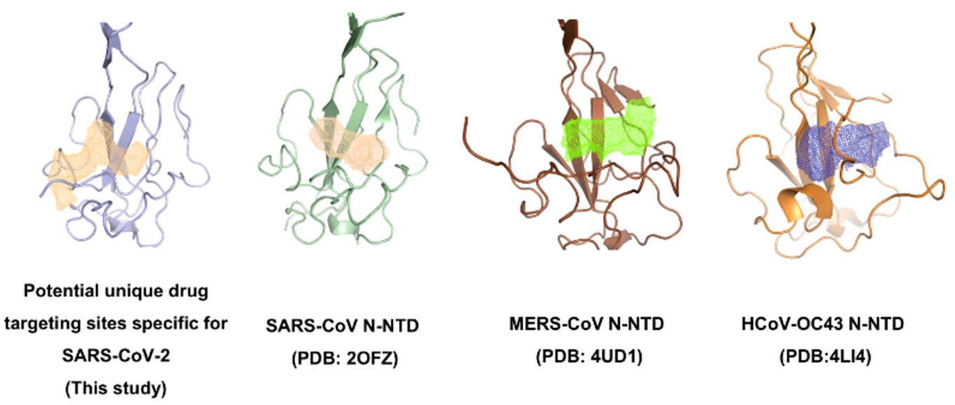
Structure of RBD NP
(2020 pmid: 32363136) 2.7 Å CoV-2. -structure is similar other RBD N-terminal domain, but surface electrostatic potential characteristics between them are distinct.
-- comparison with mild virus type HCoV-OC43 - demonstrates a unique potential RNA binding pocket alongside the β-sheet core. - vitro binding studies, - N-terminal domain, guiding the design of novel antiviral agents specific targeting to SARS-CoV-2.
RNA binding AS
(2020 pmid: 32363136) N-terminal tail residues (Asn48, Asn49, Thr50, and Ala51)-
more flexible and extended outward compared with equivalent residues in HCoV-OC43 N-NTD, possibly opening up the binding pocket into fitting with viral RNA genomic high order structure. Residues Arg89, instead of HCoV-OC43 N-NTD Tyr102, may contribute to guanosine base recognition despite the overall ribonucleotide binding that may be excluded by residues Thr55 and Ala56 in the phosphate moiety recognition site. In a previous study in which crystal structures of the CoV N-NTD in complex with five ligands (AMP, CMP, GMP, UMP, and inhibitor PJ34, respectively) were reported, Lin et al.41 demonstrated several general features for developing the hCoV-OC43 N-NTD targeting agents.
(pmid: 32511359)
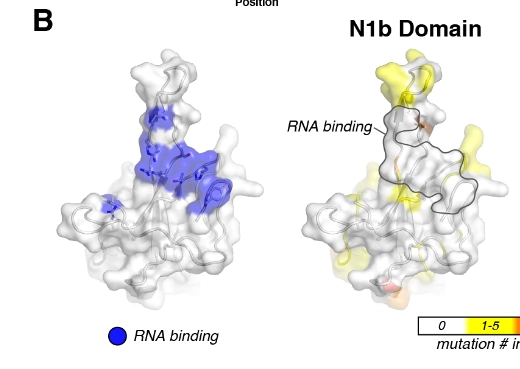
nucleotide binding in domain - was artificially done to see where the RNA binds
(2020 pmid: 32363136) HCoV-OC43 N-NTD contained adenosine monophosphate (AMP)/uridine monophosphate (UMP)/cytosine monophosphate (CMP)/guanosine monophosphate (GMP) binding site alongside the middle two β-strands of its β-sheets core41. In the complex structure of HCoV-OC43 N-NTD with ribonucleotides, the phosphate group was bound by Arg122 and Gly68 via ionic interactions, the pentose sugar ribose 2′-hydroxyl group was recognized by Ser64 and Arg164, the nitrogenous base was inserted into a hydrophobic pocket consisting of Phe57, Pro61, Tyr63, Tyr102, Tyr124, and Tyr126, mainly interacted with Tyr124 via π−π stacking forces.
- proposed that this ribonucleotide binding mechanism is essential for all coronavirus N proteins, applying to develop CoV N-NTD-target agents.
(Lin 2014 pmid: 24564608) Structure of HCoV-OC43 with nucleotide; Inhibitor of RNA binding: N-(6-oxo-5,6-dihydrophenanthridin-2-yl)(N,N-dimethylamino)acetamide hydrochloride (PJ34)
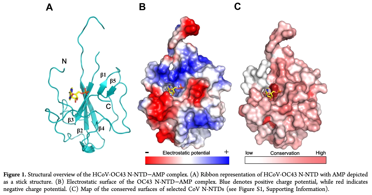
N-SARS-Cov2 RBD does not bind AMP, GMP, UMP or CMP. Binds AAGUUCGUUU - 140 microM
Regulation of RNA binding
(2020 pmid: 32363136) The crystal structures of SARS-CoV N-NTD25, infectious bronchitis virus (IBV) N-NTD26,27, HCoV-OC43 N-NTD21, and mouse hepatitis virus (MHV) N-NTD28 have been solved.
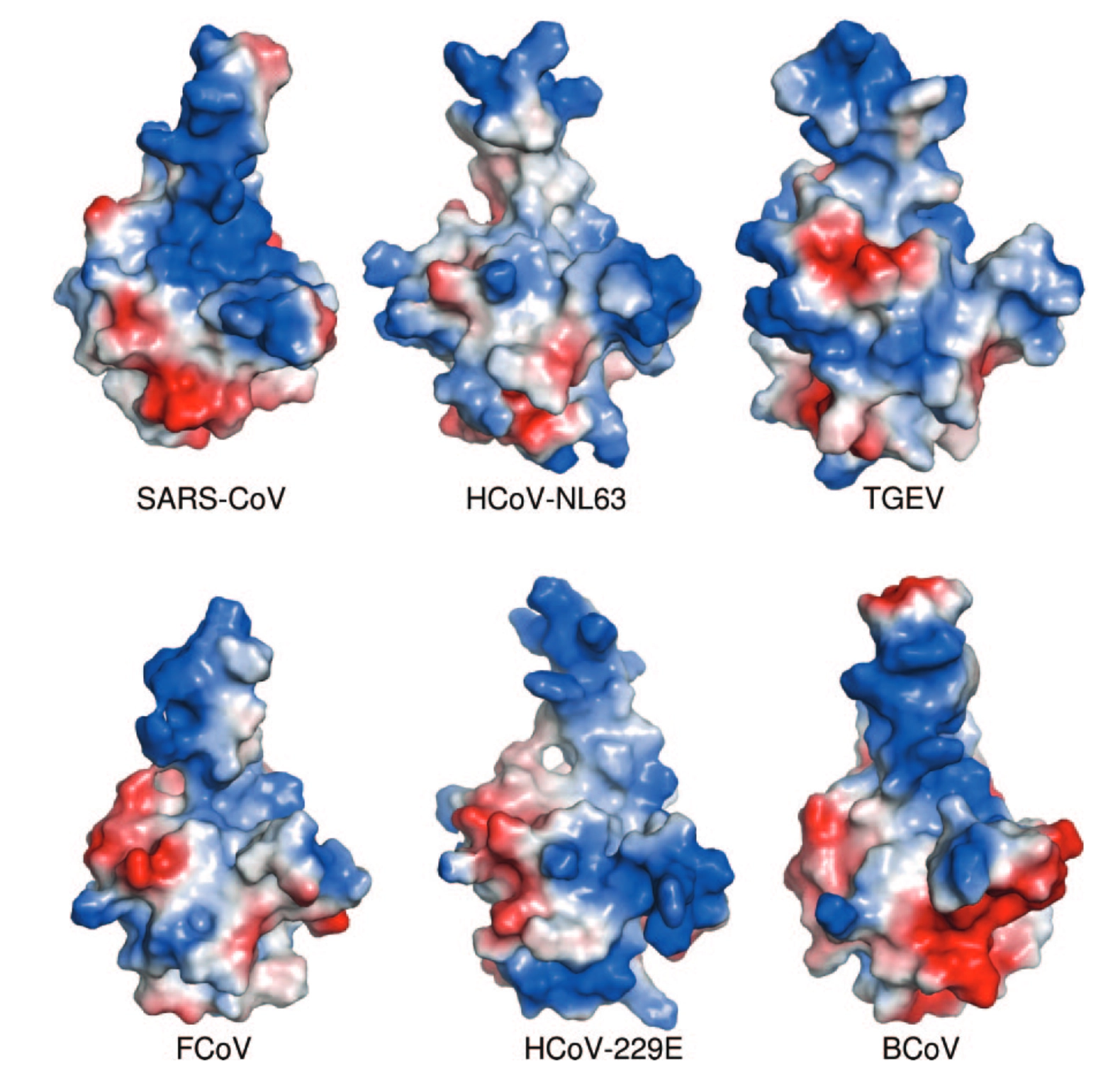
-- The CoVs N-NTD has been found to associate with the 3′ end of the viral RNA genome, possibly through electrostatic interactions. Additionally, several critical residues have been identified for RNA binding and virus infectivity in the N-terminal domain of coronavirus N proteins25,28-30.
SARS-CoV
rna binding modes
(Kuhn 2007 pmid: 17229691) comparison of crystal packing of SARS-CoV and IBV N-NTDs suggests a common mode of RNA recognition, but they probably associate differently in vivo during the formation of the ribonucleoprotein complex. - they use different modes of both RNA recognition and oligomeric assembly, perhaps explaining why their nucleocapsids have different morphologies.
Mouse Hepatitis Virus (MHV) - closely related to SARS-CoV
sequence specific binding
(Giedroc 2009 pmid: 19782089) Specifically Binds the Transcriptional Regulatory Sequence (TRS) and Melts TRS-cTRS RNA Duplexes.
- can be crosslinked to the 5′ leader RNA in infected cells,15,32
- employs aromatic amino acid nucleobase stacking interactions with a triple adenosine motif to mediate high-affinity binding to single-stranded RNAs containing the transcriptional regulatory sequence (TRS) or its complement (cTRS). Stoichiometric NTD fully unwinds a TRS-cTRS duplex that mimics a transiently formed transcription intermediate in viral subgenomic RNA synthesis.
- Mutation of the solvent-exposed Y127, positioned on the β-platform surface of our 1.75 Å structure, binds the TRS far less tightly and is severely crippled in its RNA unwinding activity. In contrast, the C-terminal domain (CTD) exhibits no RNA unwinding activity. Viruses harboring Y127A N mutation are strongly selected against and Y127A N does not support an accessory function in MHV replication.
- helix melting activity of the coronavirus N protein NTD plays a critical accessory role in subgenomic RNA synthesis and other processes requiring RNA remodeling.
- measured the binding affinity of a 5′ fluorescein (F)-labeled decanucleotide corresponding to the MHV TRS (F-5′-gAAUCUAAAC) with N219, an N domain protein fragment containing the folded NTD and the immediately adjacent intact SR-rich region (residues 60-219; Fig. S1), by fluorescence anisotropy. These data reveal that the N219-TRS complex is characterized by a Kobs = 9.0 × 10⁷ M-1 at 150 mM K+, 25 °C