NP - RBD-RNA structure - RNA binding mechanism
First description of RBD as RNA binding domain
pmid: 15147189 /2004 : NMR of RBD + binding to ssRNA (at 50uM conc NMR titrations; 16mer and 32mer of RNA )
NMR Structure CoV2 RBD + RNA
(Dinesh Boura PlosPath 2020 PMID: 33264373) additional structural DATA: 6YI3, 7ACT and 7ACS. The corresponding NMR data is available in the BMRB database under the accession code 34511.
- characterized the structure of the N-NTD and its interaction with RNA using NMR spectroscopy. Our NMR analysis is consistent with recent X-ray analysis (PDB IDs: 6M3M and 6VYO)
- NMR titrations using single-stranded and double-stranded RNA revealed a much more extensive U-shaped RNA-binding cleft lined with regularly distributed arginines and lysines. - construct hybrid atomic models of the N-NTD/RNA complex that provided detailed insight into RNA recognition.
- NMR RNA titration experiment - two ssRNA and a dsRNA. The ssRNA 7mer (5’-CUAAACG-3’) and the 10mer (5’-UCUCUAAACG-3’) - derived from the 5’ untranslated region of the genomic SARS-CoV-2 RNA containing transcriptional regulatory sequence. dsRNA random sequence (5’-CACUGAC-3’ and 5’-GUCAGUG-3’)
- 7mer and 10mer ssRNA variants affected the same N-NTD residues. The higher chemical shift perturbations observed for the 10mer oligonucleotide reflect the increased affinity towards longer RNA. The significantly perturbed residues (L56, G60, K61, K65, F66, A90, R93, I94, R95, K102, D103, L104, T165, T166, G175 and R177) formed a U-shaped binding epitope on the N-NTD surface circumventing the base of the positively charged finger.
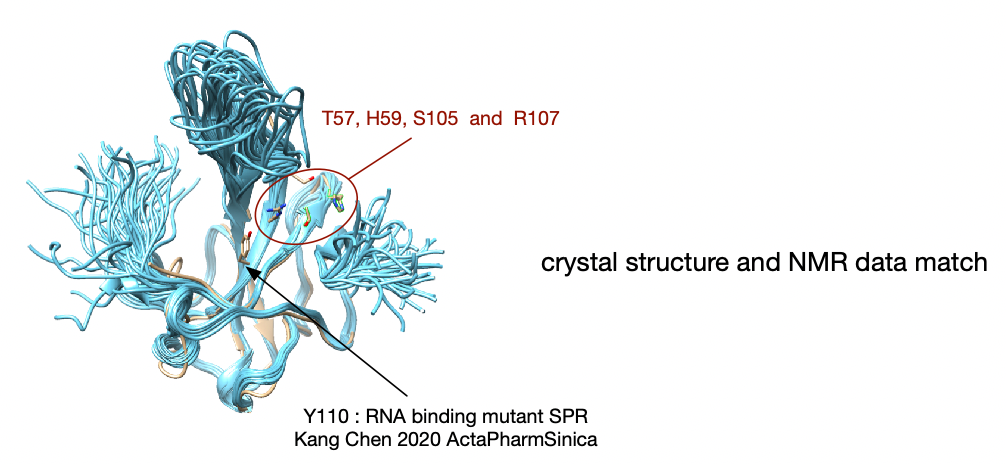
- AS perturbed by dsRNA binding. - affected residues (A50, T57, H59, R92, I94, S105, R107, R149, Y172) are located in the basic finger or close to the junction between the basic finger and the palm.
- A notable feature of the binding interface is its electrostatic potential. It is highly positive with several arginine residues (R92, R107, R149) that directly contact the RNA.
- the NMR structure revealed that the basic finger is highly flexible (Figure 2A) whereas in the crystal structures it is locked in one place by crystal lattice contacts.
- RNA binding affinity: 8uM 10nt ssRNA-hexafluorescein; binding affinity is salt dependent. see: NP - RBD Biochemical data
:-: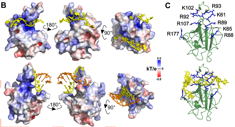
(Dinesh Boura PlosPath 2020 PMID: 33264373) additional structural DATA: 6YI3, 7ACT and 7ACS. The corresponding NMR data is available in the BMRB database under the accession code 34511.
Modeling ssRNA-N interaction
(Khan Wei 2020 CompStrucBiotech PMID: 32837710
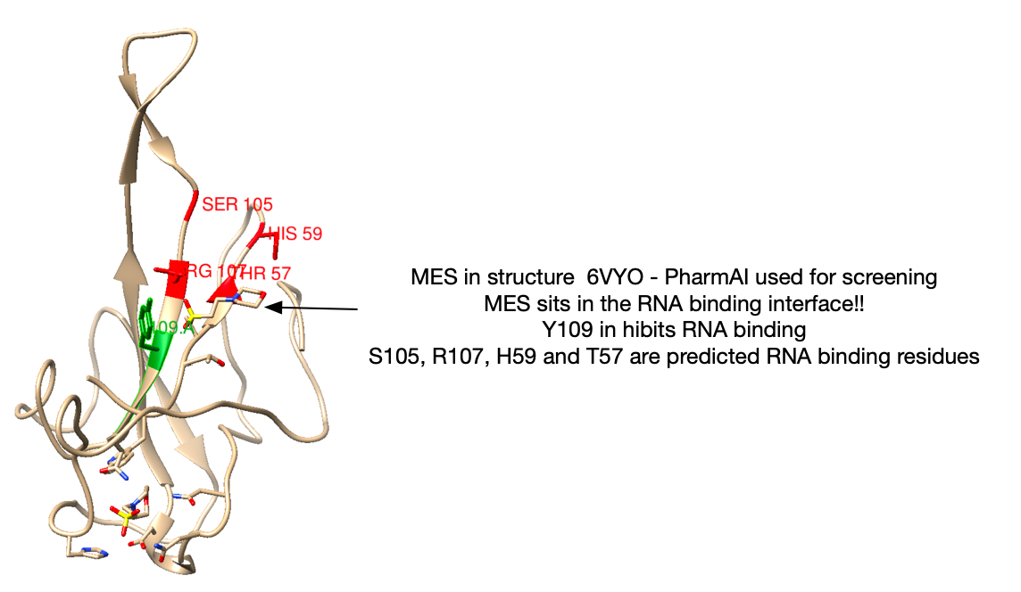
T57A, H59A, S105A, R107A, F171A, and Y172A significantly affected the dynamics and binding of RNA. Furthermore, per-residue energy decomposition analysis suggests that residues T57, H59, S105 and R107 are the key hotspots for drug discovery. - potential pharmacophores in drug designing (these residues exhibit the greatest destabilizing effects when mutated to ala).
DNAproDB server. This server mapped the interactions, and the results are shown in Fig. 1(B). Results from these analyses revealed that residues Thr57, His59, Lys61, Lys102, Asp103, Leu104, Ser105, Arg107, Lys169, Gly170, Phe171, Tyr172, Ala173, Gly175, Ser176, and Arg177 was detected, interacting with the RNA of SAR-CoV-2. Exchange of these aminoacids to alanine reduced the computational RNA binding affinity.
Ribose sugar of Adenine (A), uracil (U), and Guanine (G) formed interactions with His59, Lys61, and Tyr172. The majority of the residues were interacting with the phosphate (P) group of nucleotides (Arg107, Ala173, Gly175, Ser176, Arg177, Lys169, Gly170, and Phe171).
residues S105 and R107 are conserved among all SARS-CoV N-NTD (SARS-CoV- 2, SARS-CoV, MERS-CoV, and HCoV-OC43) [4]. Mutating S105 and R107 results in the incapability of p4a of blocking IFN production in cells infected within MERS-CoV (Siu et al. 2014).
Structure used: 6YI3 - T17, H19, S65, R67, F131, Y132
Picture: Chimera - Omnigraffle N RNA binding inhibitor
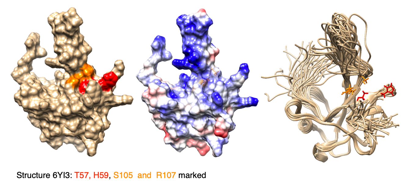
structure data used from: Dinesh Structural basis of RNA recognition by the SARS-CoV-2 nucleocapsid phosphoprotein. bioRxiv 2020. see above NMR RBD+RNA
RNA binding pocket from crystal structure
(Kang Chen 2020 ActaPharmSinica PMID: 32363136
2.7A structure of RNA-binding domain
Structure: 6M3M
Unique drug targeting pocket: From nucleotide binding structure of OC43. Arg122 and Gly68 via ionic interactions, the pentose sugar ribose 2′-hydroxyl group was recognized by Ser64 and Arg164, the nitrogenous base was inserted into a hydrophobic pocket consisting of Phe57, Pro61, Tyr63, Tyr102, Tyr124, and Tyr126, mainly interacted with Tyr124 via π−π stacking forces (Supporting Information Fig. S4). It is proposed that this ribonucleotide binding mechanism is essential for all coronavirus N proteins, applying to develop CoV N-NTD-target agents.
SPR with AAGUUCGUUU - WT) SARS-CoV-2 N-NTD is 140 µmol/L, around 2000-fold smaller than the value of Y110A mutant (KD = 330 mmol/L). This implies that WT SARS-CoV-2 N-NTD has a higher binding affinity to viral derived RNA than mutant in the binding pocket.
prediction of a druggable pocket:
PharmAI Screen
Structures used: 6WKP and 6VYO - did they look to inhibit MES binding? MES is positioned in the RNA binding pocket!!