Neuroinflammation
See also: Neuroinflammation Studies ; Inflammation & Alzheimer’s
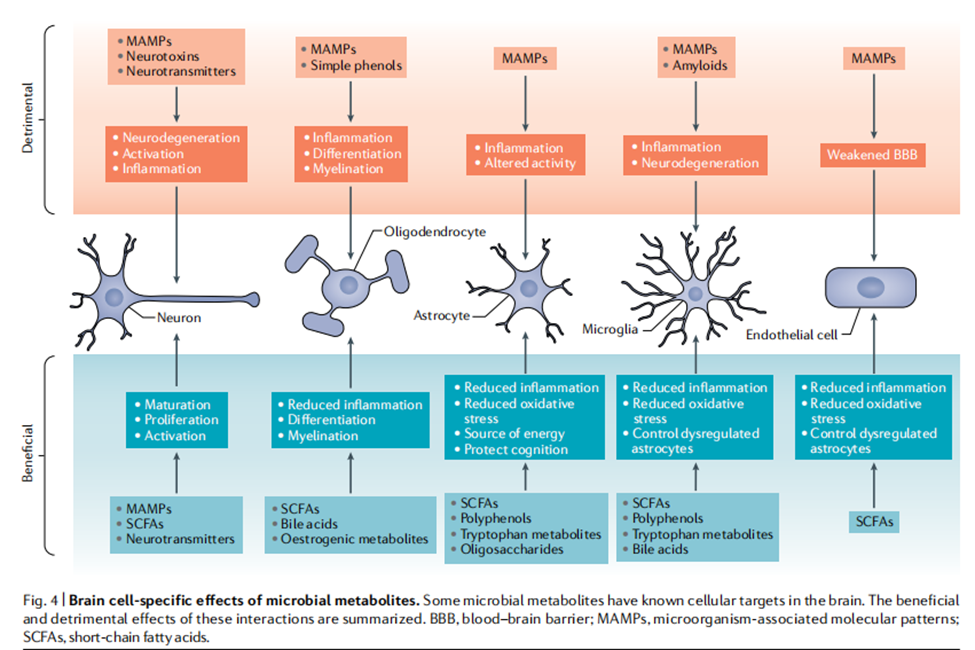
MAMPS Chart
We see on the chart below the effect of MAMPs, gram-negative microbes, and neurotoxins on neuroinflammation and neurodegeneration.
Videos
Getting to the Cause
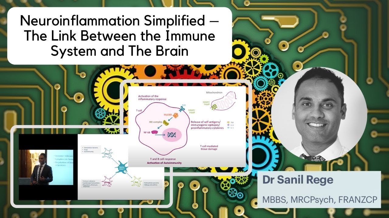
Thoughts on neuroinflammation: are persistent pathogens involved?
Neuroinflammation in Psychosis
Brain Wash: The Rise of Neuroinflammation with Dr David Perlmutter
Neuroinflammation: Causes, Assessments and Treatments with Dr Brandon Brock
Chronic Neurodeneration and Associated Neuroinflammation Following Brain and Spinal Cord Injury
Neuroinflammation Pathways
Neuroinflammation in Alzheimer's disease: current knowledge and potential directions
Centralized Pain and Neuroinflammation
Biomarkers of Neuroinflammation in Parkinson’s Disease
Overview
Neuroinflammation is inflammation of the nervous tissue. It may be initiated in response to a variety of cues, including infection, traumatic brain injury, toxic metabolites, or autoimmunity. In the central nervous system (CNS), including the brain and spinal cord, microglia are the resident innate immune cells that are activated in response to these cues. The CNS is typically an immunologically privileged site because peripheral immune cells are generally blocked by the blood–brain barrier (BBB), a specialized structure composed of astrocytes and endothelial cells. However, circulating peripheral immune cells may surpass a compromised BBB and encounter neurons and glial cells expressing major histocompatibility complex molecules, perpetuating the immune response. Although the response is initiated to protect the central nervous system from the infectious agent, the effect may be toxic and widespread inflammation as well as further migration of leukocytes through the blood–brain barrier.
Causes
Neuroinflammation is widely regarded as chronic, as opposed to acute, inflammation of the central nervous system. Acute inflammation usually follows injury to the central nervous system immediately, and is characterized by inflammatory molecules, endothelial cell activation, platelet deposition, and tissue edema. Chronic inflammation is the sustained activation of glial cells and recruitment of other immune cells into the brain. It is chronic inflammation that is typically associated with neurodegenerative diseases. Common causes of chronic neuroinflammation include:
- Toxic metabolites
- Autoimmunity
- Aging
- Microbes
- Viruses
- Traumatic brain injury
- Air pollution
- Passive smoke
Key Mechanisms
See also: Lipopolysaccharide induced neuroinflammation
Neuroinflammation is the innate immune response within the central nervous system (CNS), primarily driven by resident glial cells (especially microglia and astrocytes). It can be protective in acute settings (e.g., clearing pathogens or debris) but becomes detrimental when chronic, contributing to neuronal damage in conditions such as Alzheimer’s disease, Parkinson’s disease, multiple sclerosis, stroke, and traumatic brain injury.
Key mechanisms include the following interconnected processes:
1. Detection of Danger Signals via Pattern Recognition Receptors (PRRs)
CNS-resident cells sense pathogen-associated molecular patterns (PAMPs, e.g., bacterial LPS) or damage-associated molecular patterns (DAMPs, e.g., HMGB1, S100 proteins, misfolded proteins like Aβ or α-synuclein, ATP, mitochondrial ROS) through PRRs.
Major PRRs include:
- Toll-like receptors (TLRs), especially TLR4 (also TLR2, TLR3, TLR9).
- NOD-like receptors (NLRs), particularly those forming inflammasomes.
- Others such as RAGE and TREM2.
2. Activation of Microglia and Astrocytes
- Microglia (CNS-resident macrophages) shift from a surveillant/homeostatic state to activated phenotypes. Classically, this includes a pro-inflammatory (often called M1-like) state producing cytokines, ROS, and nitric oxide (NO), versus more anti-inflammatory/reparative (M2-like) states. Modern views emphasize a continuum of states rather than a strict binary.
- Astrocytes become reactive (astrogliosis). They can adopt neurotoxic (A1-like) or neuroprotective (A2-like) phenotypes. Microglia-derived signals (IL-1α, TNF-α, C1q) strongly drive the neurotoxic A1 state.
Microglia and astrocytes engage in reciprocal crosstalk that can amplify inflammation (feed-forward loops via cytokines and chemokines).
3. Core Intracellular Signaling Pathways
Key cascades that convert receptor activation into inflammatory gene expression include:
- NF-κB pathway: Central regulator. TLR/MyD88 signaling leads to IκB degradation, nuclear translocation of NF-κB, and transcription of pro-inflammatory genes (TNF-α, IL-1β, IL-6, iNOS, COX-2, chemokines).
- MAPK pathways (ERK, JNK, p38): Contribute to cytokine production and stress responses.
- JAK/STAT pathway: Often activated by cytokines (e.g., IL-6); promotes reactive astrocyte phenotypes and further inflammation.
- NLRP3 inflammasome: A multiprotein complex (NLRP3 + ASC + procaspase-1).
- Priming (Signal 1): Often via TLR–NF-κB, upregulates NLRP3 and pro-IL-1β/pro-IL-18.
- Activation (Signal 2): Triggered by K⁺ efflux, ROS, mitochondrial dysfunction, lysosomal damage, or protein aggregates.
- Result: Caspase-1 activation → maturation/secretion of IL-1β and IL-18, plus gasdermin D-mediated pyroptosis (inflammatory cell death).
- Other pathways: cGAS–STING (sensing cytosolic DNA/mtDNA), calcineurin/NFAT (especially in astrocytes).
4. Production and Release of Inflammatory Mediators
Activated glia release:
- Pro-inflammatory cytokines: TNF-α, IL-1β, IL-6, IL-18 (and others).
- Chemokines: CCL2, CXCL10, etc., that recruit additional cells.
- Reactive oxygen/nitrogen species (ROS/RNS): Via NADPH oxidase and iNOS, causing oxidative stress and neuronal damage.
- Other factors: Complement components, matrix metalloproteinases, ATP.
These mediators can directly harm neurons, disrupt synapses, and further activate glia.
5. Blood–Brain Barrier (BBB) Dysfunction and Peripheral Immune Involvement
Cytokines and other factors increase BBB permeability. This allows infiltration of peripheral immune cells (monocytes/macrophages, T cells—especially CD8⁺ in some contexts). Endothelial cells can upregulate MHC molecules and present antigens, amplifying T-cell responses and vascular injury.
In many neurodegenerative diseases, inflammation becomes increasingly compartmentalized within the CNS, with persistent microglial activation even after limited peripheral involvement.
6. Downstream Consequences and Feedback Loops
Chronic activation leads to:
- Neuronal damage/death (via cytokines, ROS, excitotoxicity, complement-mediated synaptic pruning).
- Impaired clearance of debris or protein aggregates.
- A vicious cycle in which damaged neurons release more DAMPs, sustaining inflammation.
Neuroinflammation is context-, stage-, and disease-dependent: early responses may be beneficial (clearance, repair), while prolonged activation is typically harmful. Therapeutic strategies often target these nodes (e.g., NLRP3 inhibitors, NF-κB modulators, microglial phenotype shifting), though translating them remains challenging.
Tissue‑State Expression of Neuroinflammation
1. Heat / Excitation
Core pattern:
Neural tissues are hot, irritated, over‑signaling, and cytokine‑driven. Microglia are in an activated, pro‑inflammatory state (M1‑skewed), producing ROS, NO, TNF‑α, IL‑1β, IL‑6.
Somatic expression:
- Burning, prickling, or “inflamed” cognitive sensation
- Irritable mood, agitation, emotional reactivity
- Photophobia, sensory overload
- Headaches with heat or pressure signatures
- Reddened or inflamed ocular tissues
Behavioral expression:
- Overthinking, rumination
- Hypervigilance
- Quick to anger or overstimulation
Tissue cues:
- Inflamed astrocytes
- Elevated oxidative stress
- NF‑κB activation
- Excitotoxic glutamate signaling
2. Wind / Tension
Core pattern:
Neural tissues are tight, spasmodic, hyperreactive, with dysregulated neurotransmission (GABA ↓, glutamate ↑). Microglia oscillate between activation and irritability.
Somatic expression:
- Tension headaches
- Jaw clenching, neck tightness
- Twitching, tremor, or spasm-like micro-movements
- Startle response heightened
- Insomnia from “wired but tired” tension
Behavioral expression:
- Anxiety with somatic tension
- Restlessness
- Difficulty settling or grounding
Tissue cues:
- Neuronal hyperexcitability
- Calcium channel dysregulation
- Stress-induced microglial priming
3. Damp / Stagnation
Core pattern:
Neural tissues are sluggish, congested, poorly clearing metabolic waste. Glymphatic flow is impaired. Cytokines accumulate. Lipid peroxidation increases.
Somatic expression:
- Brain fog
- Heaviness behind the eyes
- Slow processing speed
- Puffy or congested facial tissues
- Morning grogginess that doesn’t resolve
Behavioral expression:
- Low motivation
- Slowed cognition
- Difficulty initiating tasks
Tissue cues:
- Impaired glymphatic clearance
- Microglial debris accumulation
- Metabolic stagnation in astrocytes
4. Toxic / Constriction
Core pattern:
Neural tissues are under toxic load, constricted by oxidative stress, environmental toxins, microbial metabolites, or inflammatory debris. Microglia shift into a defensive, constricted phenotype.
Somatic expression:
- Sharp, stabbing, or “electric” pain
- Migraines with aura
- Chemical sensitivity
- Cognitive shutdown under toxic exposure
- Metallic taste or sensory distortion
Behavioral expression:
- Irritability
- Overwhelm
- Avoidance of sensory input
Tissue cues:
- ROS overload
- Mitochondrial constriction
- Lipid membrane damage
- Microglial toxin response
5. Dry / Atrophy
Core pattern:
Neural tissues are depleted, undernourished, degenerating, with reduced trophic factors (NGF, BDNF), mitochondrial decline, and chronic low-grade inflammation.
Somatic expression:
- Memory decline
- Reduced sensory acuity
- Slow synaptic response
- Dry eyes, dry mucosa
- Fatigue that feels “deep”
Behavioral expression:
- Apathy
- Reduced curiosity
- Difficulty forming new memories
Tissue cues:
- β‑amyloid accumulation
- Mitochondrial atrophy
- Reduced antioxidant enzymes
- Chronic microglial priming
Here are Assessment Notes (facilitator‑ready diagnostic cues) for Neuroinflammation, written in your Ozark Herbal Ecology style: sensory‑anchored, pattern‑driven, and immediately usable in teaching or intake.
I’m giving you clean, scannable cues organized by tissue state so facilitators can rapidly identify neuroinflammatory patterns in learners, clients, or case studies.
Assessment Notes — Neuroinflammation (Diagnostic Cues)
🌡️ HEAT / EXCITATION — Neuroinflammatory Irritation
Primary cues:
- Reports of burning, prickling, or inflamed sensations in the head or behind the eyes
- Irritability, agitation, emotional volatility
- Sensory overload: bright lights, loud sounds, crowded spaces feel “too much”
- Hot headaches, pressure headaches, or migraines with heat signatures
- Red, irritated eyes or facial flushing
- Rapid cognitive firing that feels “inflamed,” not productive
- History of cytokine‑driven flares (illness, allergies, stress)
Flags:
- NF‑κB‑style symptom clusters
- ROS-driven irritability
- Overactive microglial tone
- “Inflamed thinking” or “hot cognition”
🌬️ WIND / TENSION — Neuroreactive Tightness
Primary cues:
- Tension headaches, jaw clenching, neck tightness
- Twitching, tremors, micro-spasms, eyelid flutter
- Startle response heightened; jumpy or reactive
- Insomnia from “wired but tired” neural tension
- Anxiety with strong somatic components (tight chest, buzzing limbs)
- Difficulty settling, grounding, or downshifting
- Cognitive fragmentation under stress
Flags:
- GABA depletion / glutamate excess patterns
- Stress-induced microglial priming
- Neuromuscular tension overlaying cognitive tension
🌫️ DAMP / STAGNATION — Neurocognitive Congestion
Primary cues:
- Brain fog, heaviness, slowed processing
- Puffy or congested facial tissues, especially around eyes
- Morning grogginess that persists past waking
- Difficulty initiating tasks; “sticky” motivation
- Cognitive slowness after meals or in humid weather
- Feeling mentally “swollen” or “waterlogged”
- History of poor sleep quality or glymphatic impairment
Flags:
- Impaired glymphatic clearance
- Metabolic stagnation in astrocytes
- Cytokine accumulation without acute heat
☣️ TOXIC / CONSTRICTION — Neurotoxic Stress
Primary cues:
- Sharp, electric, stabbing pains
- Migraines with aura or sensory distortion
- Chemical sensitivity (cleaners, perfumes, smoke)
- Cognitive shutdown in toxic environments
- Metallic taste, odd sefnsory shifts
- Overwhelm from minimal stimuli
- History of toxin exposure (mold, solvents, heavy metals, chronic inflammation)
Flags:
- ROS overload
- Mitochondrial constriction
- Lipid membrane damage
- Microglial toxin-response phenotype
🌵 DRY / ATROPHY — Neurodegenerative Drift
Primary cues:
- Memory decline, difficulty forming new memories
- Reduced sensory acuity (hearing, smell, visual sharpness)
- Slow synaptic response; delayed recall
- Dry eyes, dry mucosa, “dry brain” feeling
- Deep fatigue not improved by rest
- Apathy, reduced curiosity, cognitive flattening
- History of chronic inflammation, aging, or oxidative depletion
flags:
- Low NGF / BDNF
- β‑amyloid accumulation
- Mitochondrial atrophy
- Chronic microglial priming without acute flare
Prevention/Treatment
See also: Anti-Inflammatory Alkaloids, Neuroinflammatory modulation
{Polyphenols found in fruits and vegetables are beginning to receive increased attention due to their vital role in protecting neural cells from oxidative stress and neuroinflammation associated with normal aging and chronic age-related diseases. Propolis is one of the most abundant sources of polyphenols (mainly flavonoids and phenolic acids)
Herbs with Strong Evidence for Neuroinflammation Modulation
1. Reishi (Ganoderma lucidum)
Key actions:
- Downregulates NF‑κB, TLR4, TNF‑α, IL‑1β
- Reduces microglial activation
- Enhances antioxidant enzymes
Tissue states: Heat/Excitation, Wind/Tension, Collapse
Notes: One of the most reliable neuroimmune modulators.
2. Lion’s Mane (Hericium erinaceus)
Key actions:
- Promotes NGF & BDNF
- Reduces microglial activation
- Decreases amyloid‑driven inflammation
Tissue states: Dry/Atrophy, Wind/Tension
Notes: Neurotrophic + anti‑inflammatory synergy.
3. Bacopa (Bacopa monnieri)
Key actions:
- Suppresses NF‑κB
- Reduces oxidative stress
- Protects against β‑amyloid neurotoxicity
Tissue states: Heat/Excitation, Wind/Tension, Dry/Atrophy
Notes: Excellent for excitatory neuroinflammation.
4. Gotu Kola (Centella asiatica)
Key actions:
- Inhibits TNF‑α, IL‑6, IL‑1β
- Enhances synaptic plasticity
- Reduces oxidative neuroinflammation
Tissue states: Heat/Excitation, Dry/Atrophy
Notes: Gentle but reliable neurocalming.
5. Turmeric / Curcumin (Curcuma longa)
Key actions:
- Strong NF‑κB inhibition
- Reduces microglial activation
- Protects against excitotoxicity
Tissue states: Heat/Excitation, Toxic/Constriction
Notes: One of the most potent anti‑inflammatory botanicals.
6. Green Tea (Camellia sinensis) — EGCG
Key actions:
- Inhibits microglial activation
- Reduces ROS-driven neuroinflammation
- Modulates MAPK pathways
Tissue states: Heat/Excitation, Dry/Atrophy
Notes: Excellent antioxidant neuroprotective.
7. Schisandra (Schisandra chinensis)
Key actions:
- Modulates HPA axis
- Reduces neuroinflammatory cytokines
- Protects astrocytes
Tissue states: Wind/Tension, Collapse
Notes: Adaptogenic neuroregulator.
8. Tulsi (Ocimum sanctum)
Key actions:
- Reduces IL‑6, TNF‑α, CRP
- Antioxidant + neurocalming
Tissue states: Heat/Excitation, Wind/Tension
Notes: Gentle but broad-spectrum.
9. Rhodiola (Rhodiola rosea)
Key actions:
- Reduces stress-induced neuroinflammation
- Modulates microglial reactivity
Tissue states: Wind/Tension, Collapse
Notes: Excellent for neuroinflammation driven by stress.
10. Indigo / Indole-rich plants (Crucifers, Ferments, Aristotelia)
Key actions:
- AhR modulation
- Microglial calming
- NF‑κB suppression
Tissue states: Heat/Excitation, Toxic/Constriction
Notes: You already use these in your Indole Alkaloid → Neuroinflammation card.
11. Boswellia (Boswellia serrata)
Key actions:
- Inhibits 5‑LOX, TNF‑α, IL‑1β
- Reduces neuroinflammatory edema
Tissue states: Heat/Excitation, Damp/Stagnation
Notes: Strong anti-inflammatory resin.
12. Ashwagandha (Withania somnifera)
Key actions:
- Reduces neuroinflammatory cytokines
- Protects neurons from stress-induced inflammation
Tissue states: Wind/Tension, Collapse
Notes: Excellent for neuroimmune stress patterns.
13. Ginkgo (Ginkgo biloba)
Key actions:
- Reduces microglial activation
- Enhances cerebral perfusion
- Antioxidant neuroprotection
Tissue states: Dry/Atrophy, Wind/Tension
Notes: Especially useful in vascular-driven neuroinflammation.
14. Rosemary (Rosmarinus officinalis)
Key actions:
- Inhibits COX‑2, NF‑κB
- Reduces oxidative neuroinflammation
Tissue states: Heat/Excitation, Damp/Stagnation
Notes: Underused but potent.
15. Ginger (Zingiber officinale)
Key actions:
- Inhibits TNF‑α, IL‑1β, IL‑6
- Reduces neuroinflammatory oxidative stress
Tissue states: Damp/Stagnation, Heat/Excitation
Notes: Excellent circulatory mover for stagnant neuroinflammation.
Here are Intervention Notes (herbal strategies mapped to each tissue‑state subtype) for Neuroinflammation, written in your facilitator‑ready Ozark Herbal Ecology style. These are pattern‑based, non‑clinical, and non‑directive—they describe herbal actions and strategies without giving treatment instructions.
Intervention Notes — Neuroinflammation (Herbal Strategies by Tissue State)
These are designed so you can quickly match pattern → strategy → herb category during case‑mapping.
🌡️ HEAT / EXCITATION
Pattern: Inflamed, irritated, over‑signaling neural tissue; microglial M1 activation; cytokine excess.
Herbal Strategy
- Cool inflammatory excitation
- Reduce NF‑κB signaling
- Quench ROS and oxidative heat
- Soothe irritated neural tissue
- Support microglial shift toward resolution
Herb Categories
- Anti‑inflammatories: Turmeric/Curcumin, Boswellia
- Neurocalming antioxidants: Green tea (EGCG), Bacopa
- Cooling nervines: Gotu kola, Tulsi
- Medicinal mushrooms: Reishi, Lion’s Mane
Pattern Notes
Use herbs that cool, calm, and reduce inflammatory signaling. Avoid overstimulating adaptogens during active heat.
🌬️ WIND / TENSION
Pattern: Neural hyperreactivity, spasmodic tension, neurotransmitter imbalance (GABA ↓, glutamate ↑).
Herbal Strategy
- Relax neuromuscular tension
- Support GABAergic tone
- Reduce stress‑induced microglial priming
- Stabilize synaptic firing
- Downshift “wired but tired” patterns
Herb Categories
- Relaxing nervines: Skullcap, Lemon balm, Passionflower
- Adaptogens for tension: Ashwagandha, Schisandra
- Neurotrophic mushrooms: Lion’s Mane
- Aromatic modulators: Lavender, Chamomile
Pattern Notes
Favor herbs that soften, relax, and stabilize. Avoid drying or overly astringent herbs that increase tension.
🌫️ DAMP / STAGNATION
Pattern: Cognitive heaviness, glymphatic congestion, slow metabolic clearance, cytokine accumulation.
Herbal Strategy
- Improve glymphatic flow
- Reduce edema and congestion
- Support metabolic clearance
- Increase circulation to the head
- Lighten cognitive heaviness
Herb Categories
- Circulatory movers: Ginger, Rosemary, Ginkgo
- Metabolic regulators: Schisandra, Turmeric
- Lymphatic movers: Red clover, Cleavers
- Aromatics for stagnation: Sage, Thyme
Pattern Notes
Use herbs that move, lift, and clear. Avoid heavy tonics that worsen Damp.
☣️ TOXIC / CONSTRICTION
Pattern: ROS overload, toxin‑driven microglial activation, mitochondrial constriction, chemical sensitivity.
Herbal Strategy
- Increase antioxidant capacity
- Support detox pathways (non‑directive, general)
- Protect lipid membranes
- Reduce toxin‑triggered microglial activation
- Buffer oxidative constriction
Herb Categories
- Antioxidant-rich herbs: Green tea (EGCG), Rosemary
- Medicinal mushrooms: Reishi, Chaga
- Indole/AhR modulators: Cruciferous greens, Tulsi
- Membrane protectors: Turmeric, Gotu kola
Pattern Notes
Favor herbs that buffer, protect, and neutralize oxidative stress. Avoid stimulating herbs that increase metabolic strain.
🌵 DRY / ATROPHY
Pattern: Degenerative drift, low trophic factors (NGF/BDNF), mitochondrial decline, chronic low-grade inflammation.
Herbal Strategy
- Support neurotrophic factors
- Enhance mitochondrial resilience
- Increase antioxidant enzymes
- Protect against degenerative inflammation
- Nourish depleted neural tissues
Herb Categories
- Neurotrophic herbs: Lion’s Mane, Bacopa
- Mitochondrial supporters: Rhodiola, Schisandra
- Restorative tonics: Gotu kola, Ashwagandha
- Antioxidant-rich herbs: Green tea, Turmeric
Pattern Notes
Use herbs that nourish, restore, and protect. Avoid drying astringents that worsen atrophy.
Articles/Studies
Recent studies on protective effects of walnuts against neuroinflammation.
Acacetin attenuates neuroinflammation.
Withania somnifera leaves as a potential anti-neuroinflammatory agent: a mechanistic study
Cannabidiol alleviates neuroinflammation and attenuates neuropathic pain.