NP - CTD crystal/cryo structures
CTD crystal structure w/o RNA
(Peng 2020 Embo pmid: 32914439) Structure: 7CE0
(Ye Corbett ProteinSci 2020 pmid: 32654247) PDB ID 6WZQ ; 1.4A
Crystal structure of the NP CTD +/-RNA
(Zinzula Baumeister BBRC 2020 PMID: 33039147)
boundaries of the SARS-CoV-2 N CTD to residues Thr247 and Pro 374
Crystal structures: high resolution structures at 1.44 Å (form I, PDB: 6YUN) and 1.36 Å (form II, PDB: 6ZCO) - dimeric, rhombus-shaped tile made up by the association of two CTD monomers
CTD dimerisation
Dimerization takes place via domain swapping of the two b1-b2b1 b2 hairpins, resulting in a central four-stranded bb-sheet, which is further stabilized at its edges by the a6 helix of the opposite monomer, and in the middle by the interaction between the a4 of one CTD and the a5 of the opposite one
nanoDSF of CTD
monitoring the SARS-CoV-2 N CTD intrinsic fluorescence - we observed two transitions - Tm values of 49.3 ± 0.1 °C and 54.9 ± 0.1 °C (Fig. 2E and C). - SARS-CoV-2 N CTD exists in a stable dimeric state in solution and suggest that the overall stability of the protein depends on the self-association of this domain.
RNA binding by the CTD
the putative RNA binding site that mapped to residues 248e280 in the SARS-CoV N CTD [21,33] would be conserved in SARS-CoV-2 N. Similarly, residues identified as the secondary RNA binding site in the SARS-CoV N CTD [33] correspond to Arg 319, Thr334 and Ala 336 along the b1-b2 b1 b2 hairpin (Fig. 3D). Reflecting these locations, analysis of the surface electrostatic po- tential shows that positively charged residues belonging to these amino acid tracts all cluster at opposite edges of a basic groove running transversely with respect to the dimeric interface line (Fig. 3E).
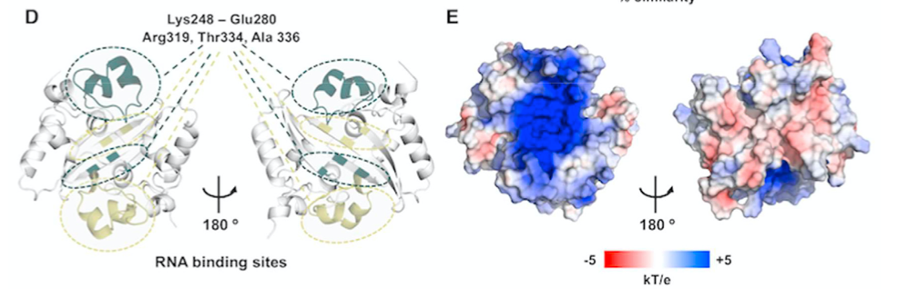
CTD is able to bind surrogates of its natural genomic substrate such as ssRNA, ssDNA, dsRNA or dsDNA, and displays micromolar binding affinities for nucleic acid moieties as short as 10 bp irrespective of the sequence [21,33,44].
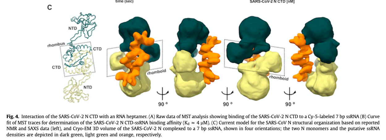
N CTD protomers pack into a helical core around which genomic ssRNA twists, so that each CTD accommodates in its positively charged groove a ssRNA tract of seven bases [12].
MST, using a 7 bp-long Cy5-fluorolabeled ssRNA probe whose sequence corresponds to the initiation of the SARS-CoV-2 genome (5’ e AUUAAAG e 3’). The SARS-CoV-2 N CTD was able to bind this probe with micromolar affinity (Kd 1⁄4 4.2 ± 0.1 mM), which is tighter than the interaction of the isolated N CTD from the closely related SARS-CoV with similar substrates (Fig. 4A and B). This result indicates that a short oligo- nucleotide of biologically relevant viral sequence can be efficiently bound by the CTD of SARS-CoV-2 N in the absence of other do- mains, and suggests that ssRNA molecules as short as 7 bp may well be cradled by the N CTD basic groove.
this is full length NP-RNA Cryo
Dimerisation domain
(Peng 2020 Embo pmid: 32914439) The dimer interface buries a surface area of 2,590 Å2. Extensive hydrogen bond interactions between the two hairpins and hydrophobic interactions between the b-sheet and the alpha helices make the dimeric structure highly stable