Podocytes
Return to Urinary System Menu
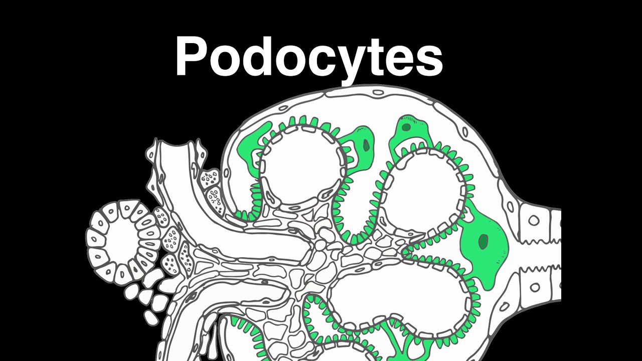
Podocytes are specialized epithelial cells that cover the outer surfaces of glomerular capillaries in the kidneys. They are also known as visceral epithelial cells.
Podocytes have a cell body with many extensions called foot processes. These foot processes are connected by unique cell junctions called slit diaphragms.
Podocytes are part of the epithelial lining of Bowman's capsule, the third layer through which blood is filtered. They maintain filtration slits, also known as slit pores, which filter blood. This filtration method is called ultrafiltration.
Podocytes play a crucial role in maintaining the glomerular filtration barrier. Loss of podocytes can lead to glomerular disease. Podocyte injury can cause proteinuria and detachment from the glomerular basement membrane
Videos
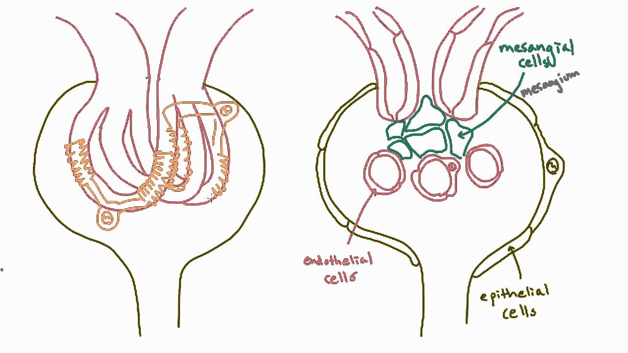
3 dimensional architecture
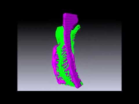
Ultrafiltration
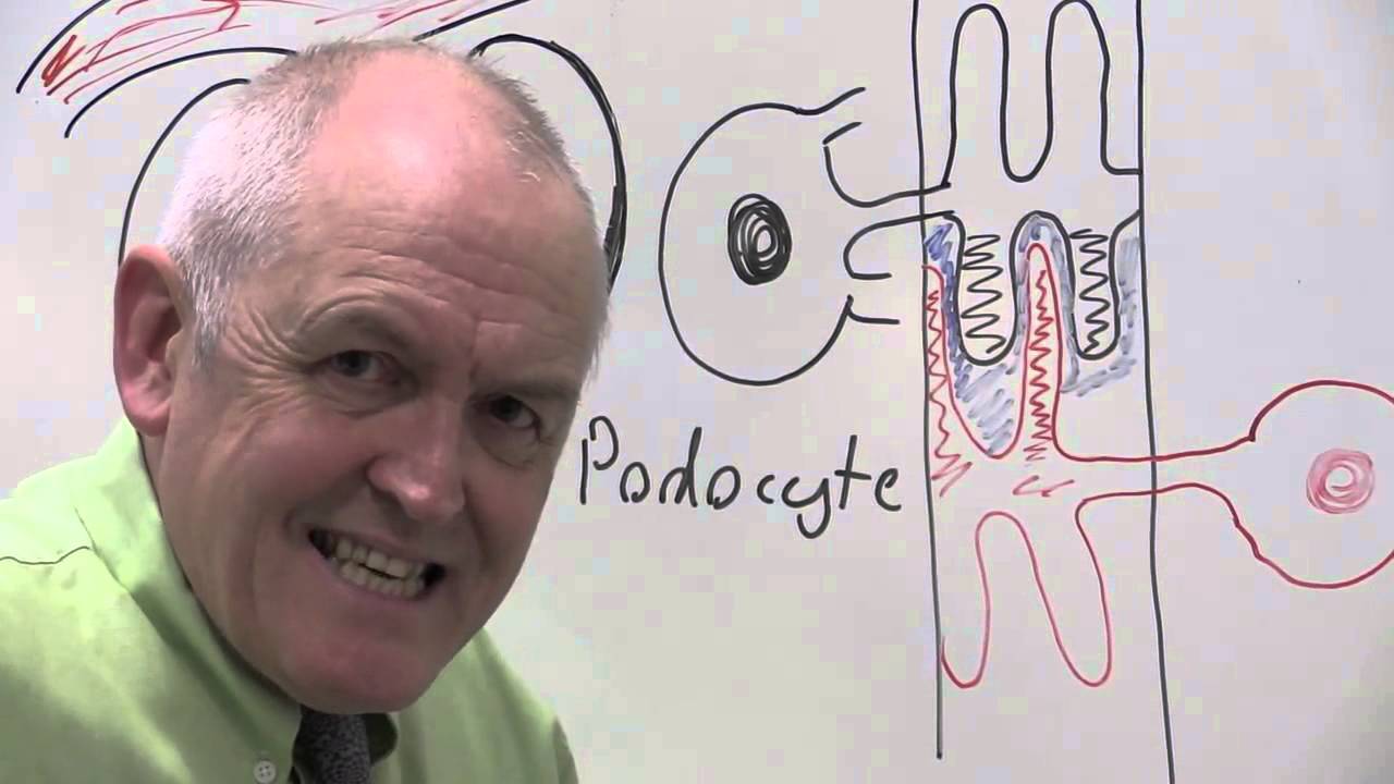
Podocyte Injury

Podocytes are specialized cells that play a crucial role in the kidney's filtration system. These cells are an integral part of the glomerulus, which is the network of capillaries in the kidney responsible for filtering blood to form urine. Podocytes are essential for maintaining the kidney's filtration barrier, and their dysfunction can lead to severe kidney diseases.
Structure and Function of Podocytes
Podocytes are unique in their structure, characterized by a cell body with numerous foot-like extensions called foot processes. These foot processes interdigitate with those of neighboring podocytes, creating a complex network that covers the glomerular capillaries. The spaces between these foot processes are known as slit diaphragms, which are critical for the selective permeability of the filtration barrier.
Key Functions
- Filtration Barrier: Podocytes form a crucial part of the glomerular filtration barrier, which prevents the passage of large molecules like proteins and cells from the blood into the urine while allowing smaller molecules like water, ions, and waste products to pass through.
- Structural Support: The intricate network of foot processes provides structural support to the glomerular capillaries, maintaining the integrity of the filtration barrier under varying blood pressures.
- Signal Transduction: Podocytes are involved in signal transduction processes that regulate their function and response to injury. They can adjust their structure and function in response to changes in the glomerular environment.
Podocyte Injury and Disease
Podocyte injury is a common feature in many kidney diseases, often leading to proteinuria (the presence of excess proteins in urine) and progressive kidney damage. The loss or dysfunction of podocytes can compromise the filtration barrier, allowing proteins and other large molecules to leak into the urine.
Common Diseases Involving Podocyte Injury
- Focal Segmental Glomerulosclerosis (FSGS): A disease characterized by scarring in the glomeruli, often associated with podocyte injury and loss.
- Minimal Change Disease (MCD): A condition where podocyte foot processes are effaced, leading to nephrotic syndrome, characterized by severe proteinuria.
- Diabetic Nephropathy: A complication of diabetes that involves podocyte damage and loss, contributing to kidney failure.
- Membranous Nephropathy: An autoimmune disease where antibodies attack podocytes, leading to thickening of the glomerular basement membrane and proteinuria.
Mechanisms of Podocyte Injury
Podocyte injury can result from various mechanisms, including:
- Immune-Mediated Damage: Autoimmune responses can target podocytes, leading to their dysfunction and loss.
- Metabolic Stress: Conditions like diabetes can cause metabolic stress on podocytes, leading to their damage and apoptosis.
- Genetic Mutations: Mutations in genes encoding podocyte proteins can lead to congenital nephrotic syndrome and other hereditary kidney diseases.
- Mechanical Stress: Increased glomerular pressure can cause mechanical stress on podocytes, leading to their detachment and loss.
Research and Therapeutic Approaches
Understanding podocyte biology is crucial for developing therapies for kidney diseases. Current research focuses on:
- Regenerative Medicine: Exploring the potential of stem cells and regenerative approaches to replace or repair damaged podocytes.
- Targeted Therapies: Developing drugs that specifically target pathways involved in podocyte injury and repair.
- Gene Therapy: Investigating the potential of gene therapy to correct genetic defects in podocyte-related diseases.
Articles/Studies
The protective effect of Phellinus linteus decoction on podocyte injury in the kidney.
Arctigenin attenuates diabetic kidney disease through the activation of PP2A in podocytes.
Astragaloside IV prevents high glucose-induced podocyte apoptosis.
The protective effect and mechanism of catalpol on high glucose-induced podocyte injury.
Fisetin attenuates diabetic nephropathy-induced podocyte injury.
Notoginsenoside R1 can inhibit the interaction between FGF1 and VEGFA to retard podocyte apoptosis.
Leonurine ameliorates adriamycin-induced podocyte injury via suppression of oxidative stress
Morroniside can reduce oxidative stress induced podocyte apoptosis.
Quercetin attenuates podocyte apoptosis of diabetic nephropathy through targeting EGFR signaling.
The protective effect of Phellinus linteus decoction on podocyte injury in the kidney.
Gastrodin inhibits high glucose-induced inflammation, oxidative stress and apoptosis in podocytes.