Bladder
Return to Urinary System Menu
[ ]
Anatomy of the Urinary Bladder
The urinary bladder is a hollow, muscular organ located in the pelvis. It serves as a temporary reservoir for urine before it is excreted from the body. The bladder's anatomy can be divided into several key components:
1. Location and Structure
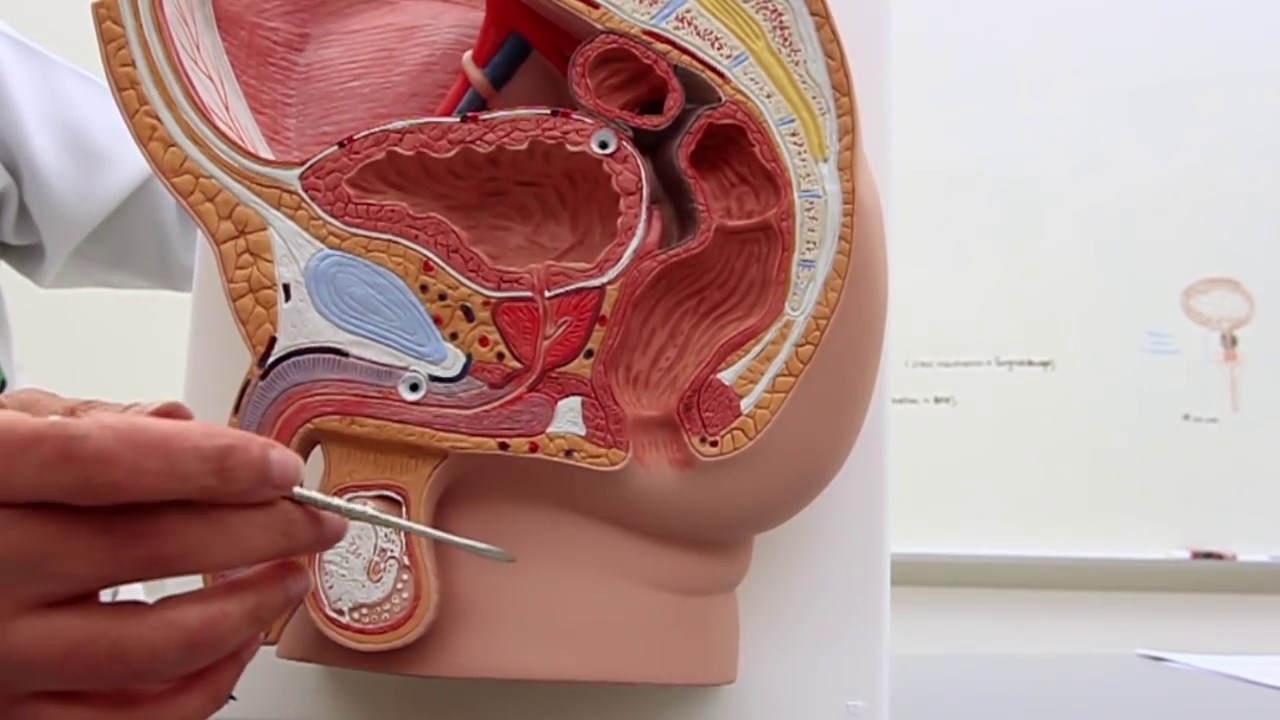
- Location: The bladder is situated in the pelvic cavity, posterior to the pubic symphysis. In males, it is anterior to the rectum, while in females, it is anterior to the vagina and inferior to the uterus.
- Shape and Size: The bladder is roughly spherical when full and collapses into a pyramidal shape when empty. Its size varies depending on the volume of urine it contains, typically holding between 400-600 milliliters in adults.
2. Layers of the Bladder Wall
The bladder wall is composed of several layers, each with distinct functions:
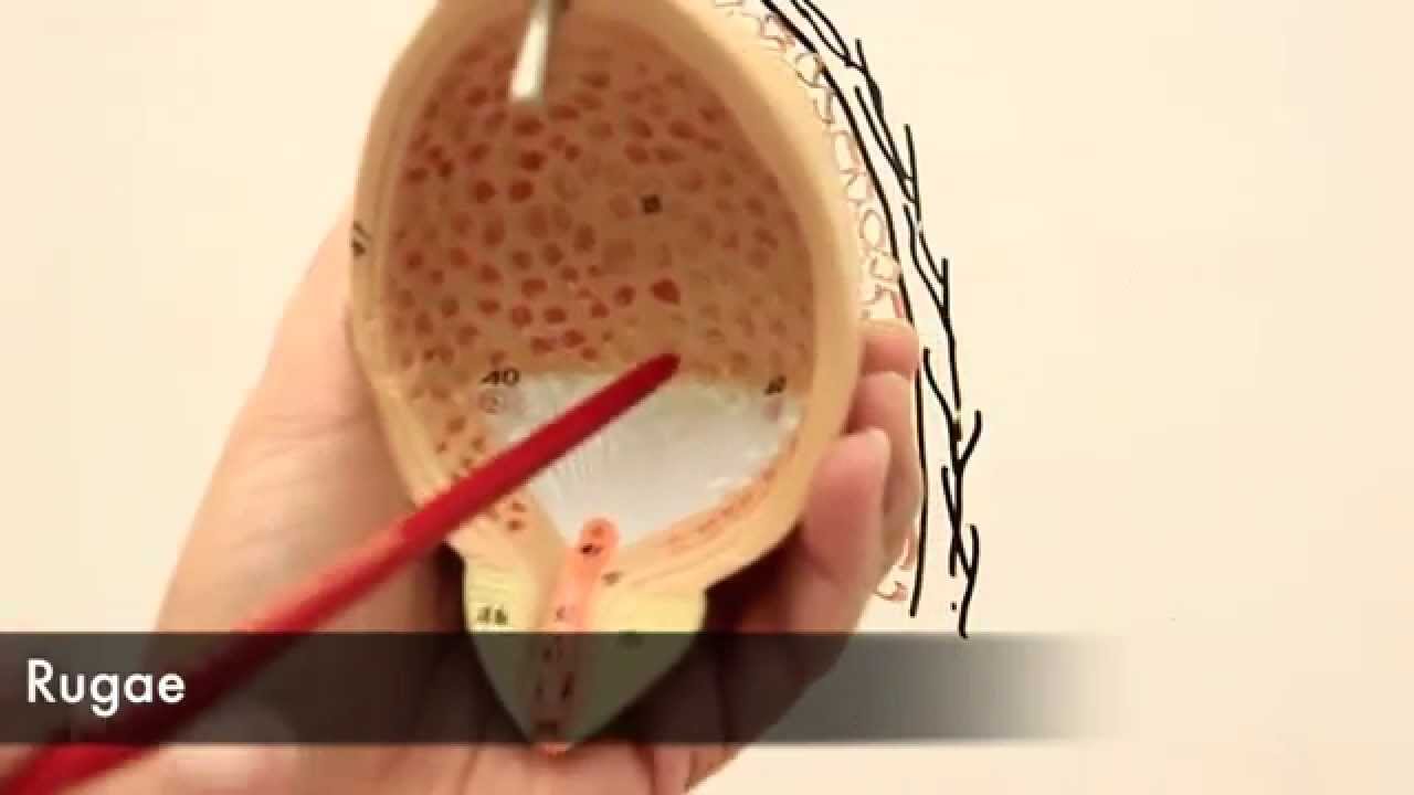
- Mucosa: The innermost layer, lined with transitional epithelium, allows the bladder to stretch as it fills.
- Submucosa: A supportive layer of connective tissue that provides elasticity.
- Detrusor Muscle: A thick layer of smooth muscle fibers responsible for the contraction of the bladder during urination.
- Adventitia/Serosa: The outermost layer, consisting of connective tissue. The superior surface is covered by the peritoneum, forming the serosa.
3. Trigone
The trigone is a triangular region at the base of the bladder, defined by the openings of the two ureters and the urethra. It is clinically significant due to its role in directing urine flow and its susceptibility to infections.
Physiology of the Urinary Bladder
The bladder's primary function is to store urine until it is convenient to release it. This process involves several physiological mechanisms:
1. Urine Storage
- Filling Phase: As urine is produced by the kidneys, it travels through the ureters and enters the bladder. The bladder's elastic walls allow it to expand and accommodate increasing volumes of urine without a significant rise in internal pressure.
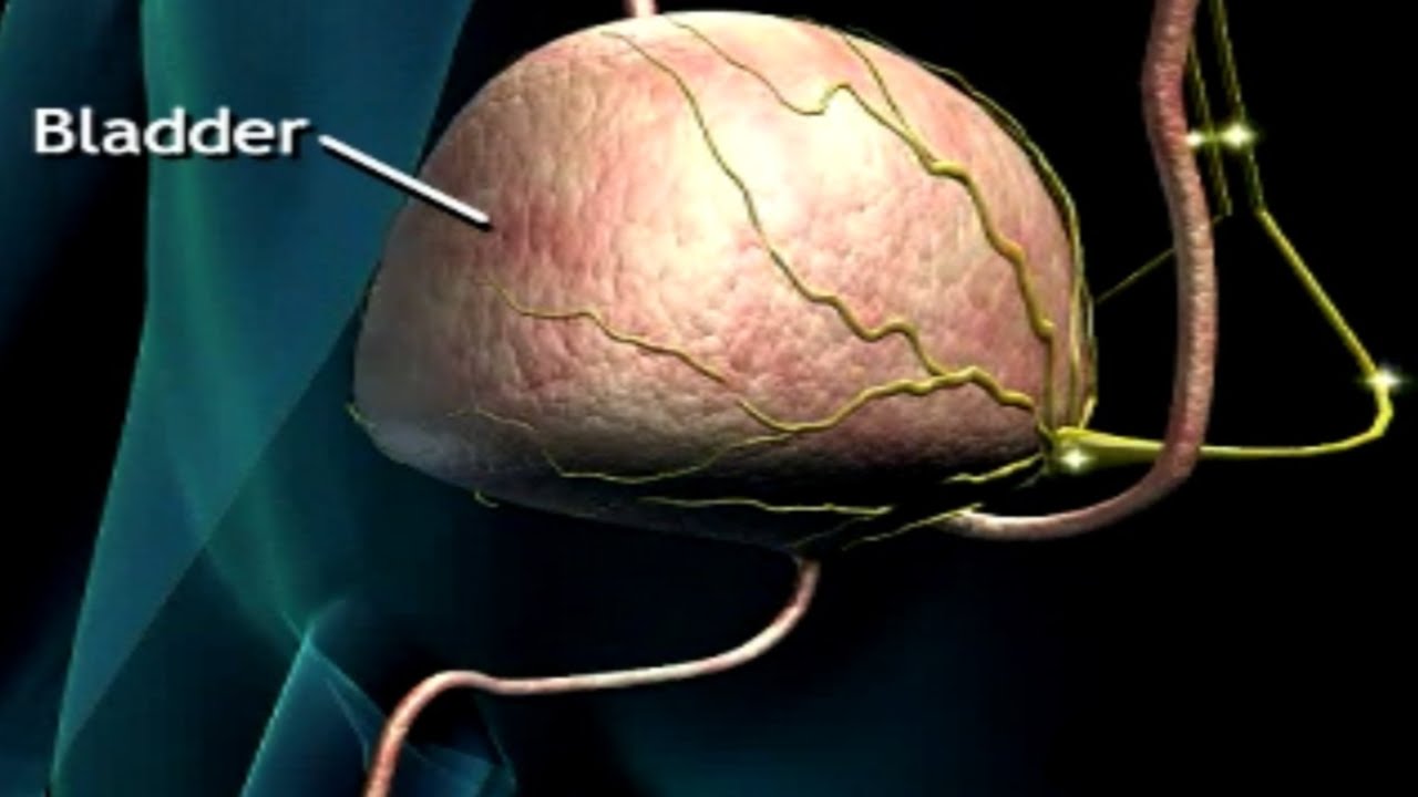
- Sensory Signals: Stretch receptors in the bladder wall detect changes in volume and send signals to the brain, creating the sensation of needing to urinate.
2. Micturition (Urination)
Micturition is the process of expelling urine from the bladder. It involves both voluntary and involuntary control:
- Involuntary Control: The micturition reflex is initiated when the bladder reaches a certain level of fullness. Parasympathetic nerves stimulate the detrusor muscle to contract, while the internal urethral sphincter relaxes.
- Voluntary Control: The external urethral sphincter, composed of skeletal muscle, is under voluntary control. This allows individuals to delay urination until it is socially appropriate.
3. Neural Control
The coordination of bladder function is regulated by the central and peripheral nervous systems:
- Central Nervous System: The pontine micturition center in the brainstem integrates sensory information and coordinates the micturition reflex.
- Peripheral Nervous System: Autonomic nerves (sympathetic and parasympathetic) and somatic nerves control the bladder and sphincters.
Clinical Significance
Understanding the anatomy and physiology of the urinary bladder is crucial for diagnosing and treating various medical conditions:
- Urinary Incontinence: A condition characterized by the involuntary leakage of urine, often due to weakened pelvic muscles or nerve damage.
- Urinary Tract Infections (UTIs): Infections that can affect the bladder, often causing inflammation and irritation.
- Bladder Cancer: A malignancy that can arise from the bladder's epithelial cells, requiring early detection and treatment.