microscope
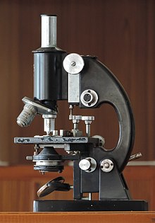
Details:
The first man to see microscopic life through a single pinhole microscope lens was [first used by] Leeuwenhoek* in 1674.
Although developed in the seventeenth century, the compound microscope only came into general use after 1830, when Joseph Lister worked out how to make lenses free of chromatic and spherical aberrations (these caused color fringes and distortion to happen). In the compound microscope a second lens enlarges the image produced by the primary lens.
By the late 19th century microscopes had reached the theoretical limit of what could be done at the visible wavelengths. In 1924 the French physicist Louis de Broglie showed that a beam of electrons could also be considered as a wave and from this idea came the electron microscope. Specimens are illuminated by an electron beam, (focused by an electromagnetic field). The most advanced form of electron microscope can magnify more than a million times.
In the 1980’s the scanning tunneling microscope was developed. Measuring the variations in an electric current generated between the microscope probe and the specimen surface, this instrument is capable of showing individual atoms.
Oneliner: The Microscope
Bibliography:
- Encyclopedia Britannica.