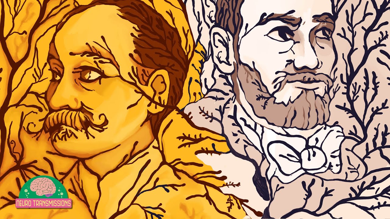
Golgi, Camillo
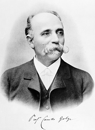
Achievements: Discovered Golgi cells in the brain.
Born: July 7, 1843/44; Corteno, Italy
Died: January 21, 1926; Pavia, Italy (Aged 83)
Field & Discipline: Medicine; Physician
Language: Italian
Education: Secondary school, Lovere (Bergamo); University of Pavia
Major Work: "Complete Works” (1903-29)
Life & Times:
At Pavia University, where he took his medical degree, Golgi worked on the pathology of the brain in the psychiatric clinic run by [worked for] Lombroso*. He graduated in 1865 with a doctoral thesis on mental diseases. He then worked at the San Matteo Hospital and assisted Lombroso at his clinic. In 1872, he was persuaded by his father to apply for an appointment as senior doctor at an institution for incurables at Abbiategrasso. Golgi came out top in the examination for the post and took up his position.
He was very disappointed in the hospital, which had few facilities, and spent much of his time to research. In the kitchen of his tiny apartment he made a laboratory and began his investigation of the central nervous organs. During this period, Golgi discovered a new method of using silver nitrate the chemical used earlier by [work related to] Fox Talbot* for his invention of paper photography. Golgi put a sample of brain-cell tissue into a silver nitrate solution and found that the silver nitrate stained the tissue.
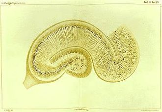
Drawing by Camillo Golgi of a hippocampus stained with the silver nitrate method.
The Spanish pathologist Ramon y Cajal wrote that when Golgi put the sample into the silver nitrate solution he saw yellow and red crystal needles forming. He removed the sample, cut it into sections and was astonished to see the nerve cells "like fine India ink drawings on transparent Japanese paper.” He could distinguish between smooth and prickly cells, and see differently-shaped cells: rods, stars and triangles.
After [influenced by work of] Perkin* produced a mauve dye from aniline in 1856, the chemical dye industry had been developed mainly by German scientists like[influenced by work of] Caro*, and German medical scientists including [influenced by work of] Ehrlich* later developed further staining techniques.
Golgi reported his conclusions in a paper in 1873. He had discovered a nerve cell later named the Golgi cell from which branches (dendrites) extended. Cajal later established the nerve cell as the basic structural unit of the nervous system. In 1874 Golgi applied the staining technique to brain pathology and was the first to describe the changes responsible for mental degeneration. He also distinguished between types of brain tumor and in 1875 published "The Morphology of the olefactory bulbs" which demonstrated that nervous impulses involve activation of the total nervous system.
Golgi was invited back to Pavia as Professor of Histology. The following year he left to become Professor of Anatomy in Siena and Turin but returned to Pavia in 1877 as an histologist. He was eventually given his own laboratory and began to train pupils. In 1880 he described the point at which sensory nerve fibres end in branchings enclosed in a tendon the site is now known as the Golgi tendon organ. He also distinguished between motor and sensitive fibres in the spinal marrow, and described the existence of two categories of cells which differ in the structure of their extensions.
In 1883 he discovered in nerve cells a network of small fibres, cavities and granules which is now known as the Golgi complex. This is found in all cells except bacteria, and may be related to the structuring of membrane, as well as to the storage and transport of materials in the brain.
After 1884, Golgi turned his attention to the study of malaria. He solved the mystery of intermittent fever attacks by describing the cycle of the plasmodium parasite which produces the disease: the onset of fever in the patient occurs when parasite spores are released by the red blood cells. He also explained the difference between tertian fever, (which occurs every other day, and quartan fever, which occurs every third day (reason: different varieties of the parasite have different cycles). His discovery made possible the correct diagnosis of malaria type in a patient, and also allowed for attacks to be predicted and medication given at the optimum time to counter the attack.
Many young scientists came to Pavia to study the new research methods relating to the nervous system. Among them was [a pal of] Nansen*, the polar explorer with whom Golgi formed a warm friendship. In 1889 he studied the structure of the kidney and demonstrated details of the development of renal structures. In 1898 he was the first to describe a small structure, now called Golgi's internal reticular apparatus, which exists in the plasma of cells.
In 1900 Golgi was elected as a senator and appointed as director of public health. He founded a laboratory for health inspection and campaigned for malaria control. He attempted to improve social hygiene among the poor. In 1903 Golgi was awarded a large sum by the Wurzburg medical faculty for his work in brain anatomy research.
He set up a new laboratory, where he worked on rabies. In 1906 he was awarded the Nobel prize jointly with Cajal. During World War I (1914-18) Golgi worked as a clinical doctor and set up a special field hospital for the treatment of soldiers with brain injuries. He retired in 1918 but continued to lecture regularly on the histology of the nervous system and to make daily visits to his laboratory.
Extra Connections: He shared the Nobel Prize with [shared Nobel Prize with] Ramon y Cajal*
Assessment: Golgi is remembered for his discovery and description of structures in the nervous system.
Oneliner: Camillo Golgi, discoverer of Golgi brain cells
Bibliography:
- Dictionary of Scientific Biography.
- Pilleri, G., “Camillo Golgi,” in Biographical Sketches published for the 50th anniversary of the Bern University Brain Anatomy Institute, Verlag des Hirnanatomischen Institutes, Ostermundigen, Switzerland, 1984.
Father: Father: Alessandro Golgi
Mother: Carolina
Siblings: 2 brothers
Spouse: Lina Aletti
Children: None
Multimedia:
Video: The Black Stain That Changed Neuroscience: